Introduction
Acne vulgaris, ordinarily known as acne, is a common skin disease that affects most adolescents and adults at some time in their lives (1). Acne is a chronic inflammatory disease of the pilosebaceous unit. The lesions are localized in “acne prone areas”, where sebaceous follicles are most common, which includes the surface skin of the face, neck, chest, and back. Acne generally develops in young people due to several factors including food, stress, the use of cosmetics, hormonal imbalance, or bacterial infection (2). It is accepted that inflammation in acne vulgaris is mainly induced by an immunological response to Cutibacterium acnes (once referred to Propionibacterium acnes) (2). This bacterium plays a critical role in the development of inflammatory acne when it overgrows and colonizes the pilosebaceous unit (3).
C. acnes is a gram-positive non-spore-forming anaerobic bacterium; it is pleomorphic and rod shaped, and can produce propionic acid. The organism forms units in the normal flora of the oral cavity, large intestine, conjunctiva, external ear canal and skin, where it dominates over other normal flora components in pilosebaceous follicles (4).
Staphylococcus aureus is a common foodborne pathogen capable of causing a variety of human infections (5). In man, S. aureus causes two main infection types: (1) cutaneous or mucosal infections and (2) septicemic infections that are generally associated with visceral (abscesses, endocarditis, lung) or bone (osteomyelitis) infections (6). S. aureus has the capacity to readily acquire antimicrobial resistance, which makes remedy difficult. Treatment of acne invloves the use of topical or systemic antibiotics; however, gradual increase in incidences of antibiotic resistance in the causative bacterium has led to a serious need for effective, non-antibiotic treatments (5).
Many researchers have worked toward the development of remedial agents for acne that do not contribute to resistance but still have high antibacterial activities (1). In recent years, there has been increasing interest in healthy lifestyles and aging using products of plant origin. A large number of aromatic, spicy, medicinal, and other plants that belong to the Asteraceae family contain chemical compounds that exhibit antimicrobial and antioxidant properties (7).
Mugwort (Artemisia) species are part of the Asteraceae family and have been used in traditional medicine to treat serious diseases, including microbial and viral infections (8). Above all, Artemisia iwayomogi Kiramura is a well-known herbal hepatotherapeutic drug that has been traditionally used in Korea and/or China over long periods of time for antitumor, immunomodulating, antimutagenic, antioxidant, antibacterial, antifungal, liver-defense and choleretic purposes (8,9). Artemisia iwayomogi has attracted substantial attention for its potential use as a chemopreventative agent (10). Pharmaceutical researchers have identified a number of terpenoid acids, polyenes, phenols, coumarins, chromones, and flavonoids as the ingredients of the crude drug. There are also numerous reports on the biological activities of Artemisia against human pathogens comprising viruses, fungi, and yeasts (11); however, there are only a few research reports that examine the effects of A. iwayomogi against C. acnes, one of the major acne-causing bacteria. Therefore, the objectives of this research are 1) to examine the antimicrobial activities of mugworts against C. acnes and S. aureus, and 2) to identify the major compounds responsible for antimicrobial activity.
Materials and Methods
Cutibacterium acnes KCTC 3314 and Staphylococcus aureus KCTC 3881 were cultured in Reinforced Clostridial Medium (Difco Laboratories, Detroit, MI, USA) and Nutrient broth (Difco Laboratories, Detroit, MI, USA), respectively. The C. acnes culture was incubated at 37℃ for 72 h under anaerobic conditions in a candle gas jar (GasPak EZ Container Systems, Franklin Lakes, N.J., USA). The S. aureus culture was incubated in an incubation shaker at 37℃ and 200 rpm for 24 h. After incubation, both cultures were washed twice with Butterfield’s phosphate buffer (BPB) by centrifuging at 5,000 rpm for 5 min at 5℃. The collected bacteria were re-suspended in BPB and the bacterial population was determined by measuring the optical density at 650 nm with a spectrophotometer (UVmini 1240, Shimadzu, Kyoto, Japan).
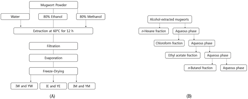
Korean native mugworts including In-jin-ssuk (Artemisia iwayomogi) and Yak-ssuk (Artemisia princeps) were purchased from a local market located in Daegu, South Korea. The leaves and stems of the mugworts were harvested in the Kangwha area of South Korea during the summer of 2012 and sun-dried immediately after harvesting. Each mugwort was ground into a fine powder prior to extraction. Ground mugwort (50 g) was separately added to 500 mL of distilled water, 500 mL of 80% ethanol, and 500 mL of 80% methanol, and stirred in a water bath at 60℃ for 12 h. Each extract was then filtered through Whatman No.2 filter paper, and the extracted residue was re-extracted twice (12). The solvent from each extract was completely removed by rotary evaporation. The extracts were freeze-dried and stored at 4℃ prior to use. The extraction yield of each sample was determined using the following equation: Extraction yield (%) = [weight of extract (g)/weight of dried mugwort (g)]×100.
The methanolic or ethanolic mugwort extracts were further fractionated using organic solvents, including n-hexane, chloroform, ethyl acetate, and n-butanol. n-Hexane (200 mL) and water were added to the alcohol extract in a separating funnel to obtain the n-hexane fraction. The n-hexane phase was separated, and the remaining aqueous phase was successively treated with various solvents. Chloroform, ethyl acetate and n-butanol fractions were obtained using similar procudures (13). The overall fractionation process is illustrated in Fig 1. Following fractionation, each solvent was completely removed by rotary evaporation and each fraction was freeze-dried and stored at 4℃ prior to further use.
The alcoholic mugwort extracts were incorporated into cultural broths at concentrations of 0, 5, 10, 15, and 20 mg/mL. Streptomycin (5 mg/mL) (Dufhefa, Biochemie, Netherlands) was used as the positive control. Test bacterial cultures were inoculated at concentrations of 106 to 108 CFU/mL, and incubated at 37℃ for 48 h while shaken at 200 rpm. An aliquot of 1 mL culture was washed twice with BPB by centrifugation at 5,000 rpm for 5 min using a centrifuge (Microcentrifuge 1730R, Labogene, Seoul, Korea). The washed bacteria were re-suspended in 1 mL of BPB, after which 100 μL of the bacterial suspension was transferred in a 96-well microplate (BioTek, Seoul, Korea), and the optical density at 650 nm was measured using a plate reader (BioTek, Seoul, Korea). The inhibitory effect of the mugwort extract was calculated using the following equation: Inhibitory Effect (%) = (O.D. measured without the mugwort extract-O.D. measured in the presence of the mugwort extract)/O.D. measured without the mugwort extract×100.
The n-hexane, chloroform, ethyl acetate, and n-butanol fractions from the alcoholic mugwort extracts were further examined to determine their minimum inhibitory concentrations (MICs). The broth containing each fraction was prepared as described in the previous section, after which the culture was immediately poured onto Petri plates containing the designated medium for each bacterium. The inoculated plates were incubated at 37℃ for 24 h or 72 h. At the end of the incubation period, the plates were evaluated for the presence or absence of growth. The MIC was determined as the lowest concentration of mugwort extract that resulted in the absence of bacterial growth.
The antimicrobial activities of the mugwort extracts were also determined using the agar diffusion method. C. acnes and S. aureus were cultivated to concentrations of approximately 106 CFU/mL. An aliquot of 1 mL of each culture was spread onto Reinforced Clostridial agar for C. acnes and nutrient agar for S. aureus. Blank paper discs of 6 mm in diameter (BBL, Cockeysville, MD, USA) were impregnated with the specified concentrations of mugwort extracts and placed on the surfaces of the agar plates. The C. acnes plates were then incubated at 37℃ for 72 h in an anaerobic jar (GasPak EZ Container Systems, Franklin Lankes, N.J., USA). The S. aureus plates were incubated at 37℃ for 24 h. Streptomycin (5 mg/mL) and BPS buffer were used as positive and negative controls, respectively. Followng incubation, the diameter of each inhibition zone was measured at two cross-sectional points, and the average was used to calculate the area of the inhibition zone. The antimicrobial activity of each mugwort extract is expressed by the square of zone width according to the following equation (14): Square of zone width (mm2) = [diameter of clear zone-diameter of the blank disc (6 mm)]/2.
Mugwort samples were extracted using 80% methanol and filtered through filter paper. The methanol was completely removed from the extract by rotary evaporation. Each residual extract was dissolved in water for further fractionation with chloroform. GC/MS was performed using a 6,890 GC gas chromatograph coupled to a 5,975 mass spectrometer (HP, Palo Alto, CA, USA). Compounds were separated on a 30 m×0.25 mm i.d. capillary column with a 0.25-μm-thick coating (15). The column temperature was maintained at 60℃ for 5 min after injection, then programmed to increase at 10℃/min to 250℃, and maintained at that temperature for 5 min. Split injection with a split ratio of 5:1 was used, and helium was used as the carrier gas at a flow rate of 1 mL/min. The spectrometer was operated over the 29-800 amu mass scan range, and a solvent delay time of 3 min was employed. The chemical constituents were identified by comparing their relative retention times and mass spectra with those obtained from authentic sample and/or the NIST/NBS and Wiley libraries spectra (9).
Each experiment was repeated three times. One way analysis of variance (ANOVA) was performed using SPSS software (version 11.5, SPSS Inc., Chicago, IL, USA). Differences among means were analyzed using Duncan’s Multiple Range test (DMRT), and the significance level was defined as p<0.05.
Results and Discussion
The extraction yields of Yak-ssuk (A. princeps) using water, ethanol, and methanol were 13.9, 9.1, and 10.8%, respectively. Similar extraction yields were also obtained for In-jin-ssuk (A. iwayomogi). The highest yield was obtained from the water extract, whereas the lowest yield was obtained from the methanol extract. Kim et al. (16) also reported that the highest extraction yield was obtained from the water extract of Hizikia fusiforme. These results indicate that the variances in extraction yields among the selected solvents are attributed to differences in solvent polarity, which results in different compounds extracted from the mugworts by the various solvents.
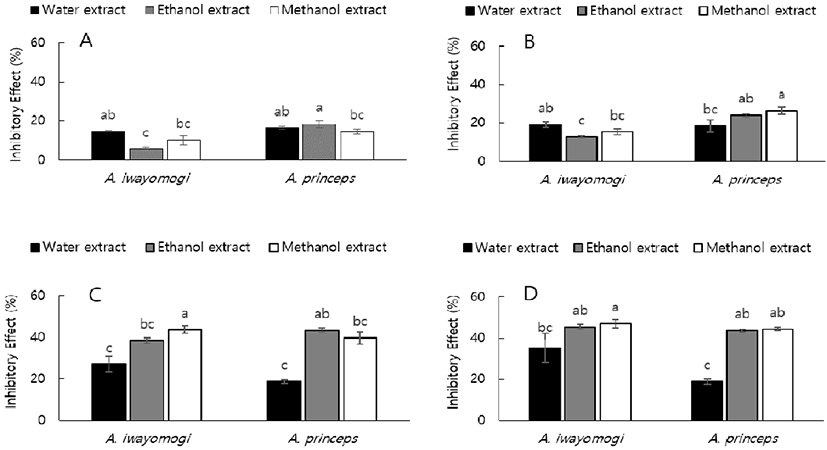
The inhibitory effects of mugwort extracts against C. acnes and S. aureus are presented in Figs. 2 and 3, respectively. The inhibitory effects of the In-jin-ssuk (A. iwayomogi) and Yak-ssuk (A. princeps) extracts were both observed to increase as the extract concentrations increased from 5 to 20 mg/mL. At 20 mg/mL, the inhibitory effects of the ethanol and methanol extracts of the two mugworts against C. acnes were significantly higher than those of the water extracts (p<0.05); however there was no significant difference between the ethanol and methanol extracts (Fig. 2D).
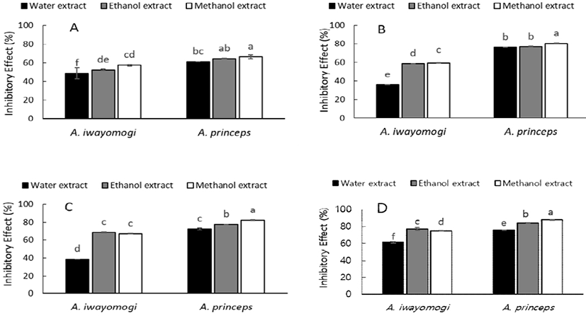
The mugwort extracts exhibited similar trends toward S. aureus to those observed for C. acnes; however a significant difference was observed between the two alcohol extracts. The ethanol extract from In-jin-ssuk exhibited the highest inhibitory effects, while the methanol extract from Yak-ssuk was most active (Fig. 3D). These results reveal that alcohols, such as ethanol or methanol, are more effective solvents for extracting antimicrobial compounds present in mugwort than water; therefore, the ethanol and methanol mugwort extracts were further fractionated.
The antimicrobial activities of both the ethanol and methanol extracts of In-jin-ssuk (A. iwayomogi) fractionated with selected solvents were further evaluated by determining their MICs, which are the lowest concentrations of the antimicrobials that inhibit the visible growth of a microorganism. Antimicrobial potency was qualitatively and quantitatively assessed using the MIC values. As presented in Table 1, fractionation of the alcohol extracts of In-jin-ssuk (A. iwayomogi) using selected solvents led to improved antimicrobial activities against acne-causing bacteria. The MIC of the chloroform fraction of the methanol extract was found to be 10 mg/mL against both C. acnes and S. aureus, while the MIC of the chloroform fraction of the ethanol extract was 15 mg/mL against both bacterial strains.
The antimicrobial activities (MICs) of the n-hexane and ethyl acetate fractions were found to be 20 mg/mL against both C. acnes and S. aureus. The MICs of the ethyl acetate and hexane soluble extracts of Eisenia bicyclis have previously been reported to be 64 μg/mL and 128 μg/mL, respectively, against C. acnes (17). The MICs of tea-tree oil were determined to be between 0.31% and 0.63% for C. acnes, and between 0.63% and 1.25% for S. aureus, indicating that tea-tree oil possess substantial antimicrobial activities against both C. acnes and S. aureus (18). Extracts of rose, duzhong, and yerba mate also exhibit notable antimicrobial activities against C. acnes (19). Among these extracts, the duzhong extract is antimicrobially most active against C. acnes with an MIC of 0.5 mg/mL, while the yerba mate extract showed moderate antibacterial activity against C. acnes with an MIC of 1 mg/mL. In addition, the in vitro antimicrobial activities of more essential oils, medicinal plants, and chemicals against C. acnes have been reported by several research groups (20-22).
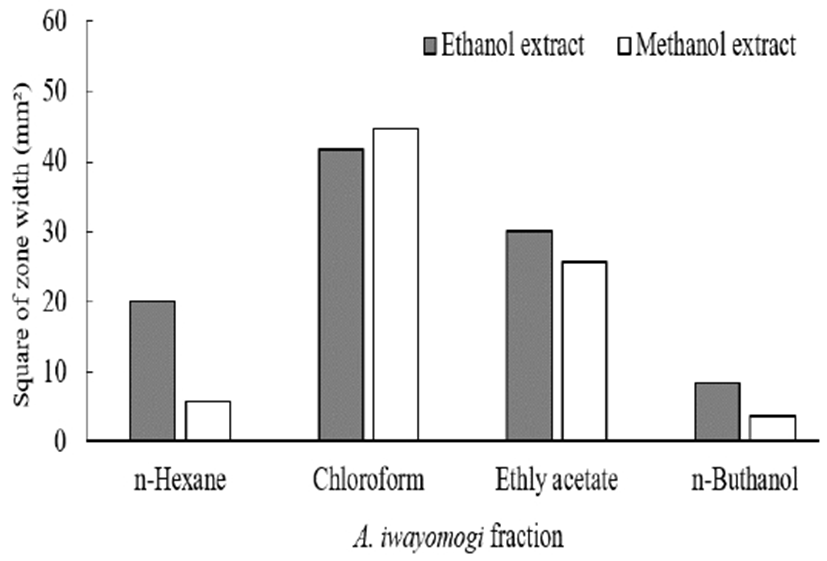
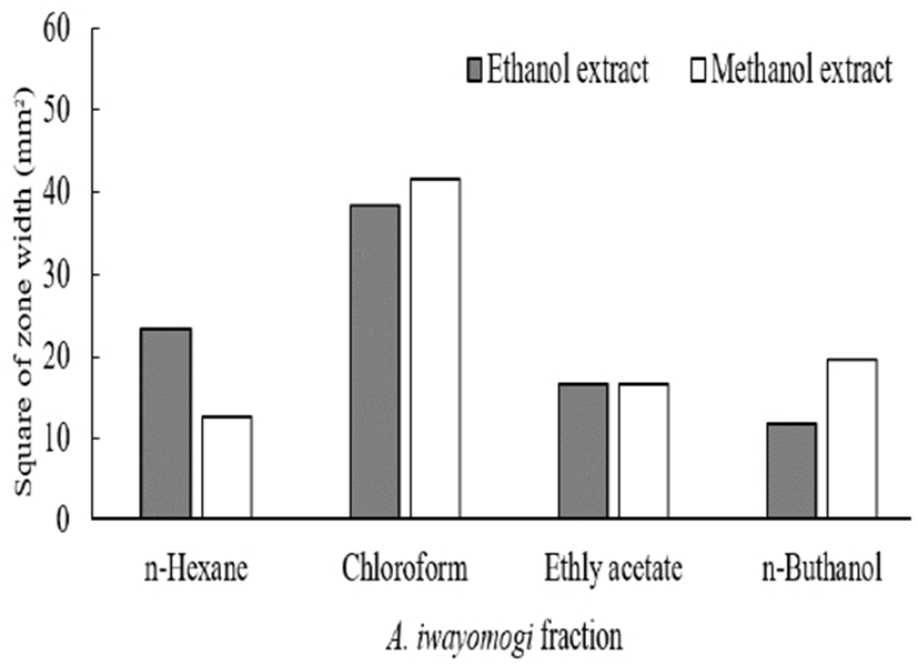
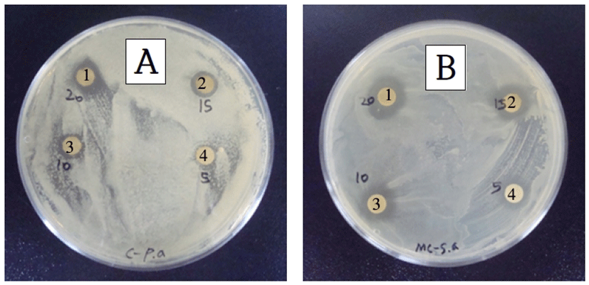
The antimicrobial potencies of the mugwort extracts were qualitatively and quantitatively assessed by the presence or absence of inhibition zones, and by the diameters of these zones. Although the alcohol extracts of In-jin-ssuk (A. iwayomogi) inhibited the growth of both C. acnes and S. aureus, these extracts did not produce distinct clear zones when tested using the disc diffusion method. Further fractionation of the alcohol extracts of In-jin-ssuk (A. iwayomogi) using selected solvents resulted in extracts with higher antimicrobial activities against both C. acnes and S. aureus; these extracts produced measurable clear zones in the disc diffusion assay. The clear zones of the solvent-fractionated In-jin-ssuk (A. iwayomogi) from the methanol extracts are presented in Fig. 4. Among the fractions, the chloroform fractions of In-jin-ssuk (A. iwayomogi) exhibited the largest clear zones against both C. acnes and S. aureus, when tested at concentration of 20 mg/mL. The square of zone width of a clear zone is often used to relate the antimicrobial activity of a compound (14). As presented in Fig. 5, the chloroform fractions from the methanol and ethanol extracts were calculated to have the largest squares of zone widths among the fractions, while the n-butanol fractions from the methanol and ethanol extracts exhibited the smallest squares of zone widths. Tea-tree oil at a concentration of 5 mL has been reported to show squares of zone widths of 78 mm2 and 230 mm2 for inhibition zones against C. acnes and S. aureus, respectively (18). The squares zone widths of the clear zones from the In-jin-ssuk (A. iwayomogi) fractions against S. aureus exhibited similar trends to those observed for C. acnes (Fig. 6). The results of a previous study showed similar results to those of the chloroform fractions of the In-jin-ssuk (A. iwayomogi) extract in this study.
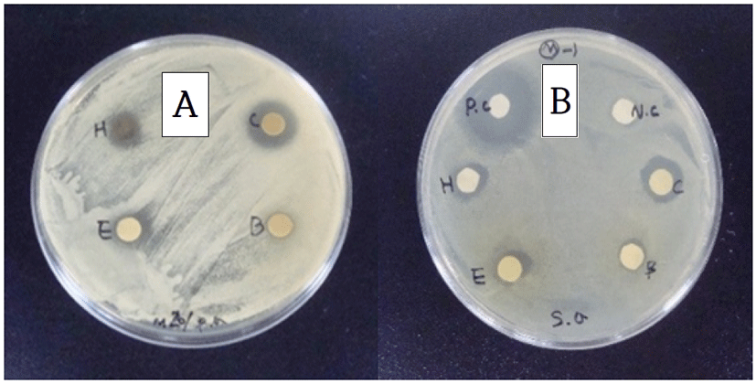
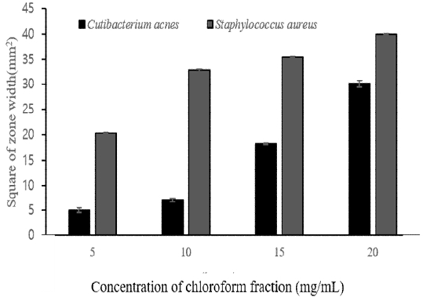
The clear-zone size of the chloroform fraction of the methanol extracted In-jin-ssuk (A. iwayomogi) was observed to increase with increasing fraction concentration (Fig. 7). The largest inhibition zone was observed at a chloroform-fraction concentration of 20 mg/mL (Fig. 8). Based on these results, the chloroform fractions from methanol extracts exhibit the strongest antimicrobial activities against C. acnes among the tested fractions with the lowest MIC of 10 mg/mL and the largest inhibition zone (12-18 mm diameter). Therefore, the chloroform fraction from the methanol extract of In-jin-ssuk (A. iwayomogi) was further analyzed by GC/MS in order to identify the compounds responsible for antimicrobial activity.
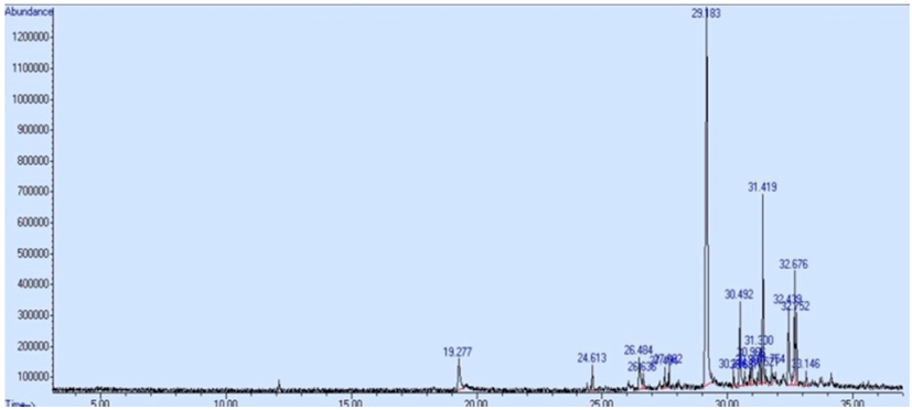
The GC/MS trace of the chloroform fraction of the In-jin-ssuk (A. iwayomogi) methanol extract is presented in Fig. 9, which reveals that scopoletin (48.39%), isofraxidin (5.49%) and 2-furanmethanol (6.12%) are main components of the In-jin-ssuk (A. iwayomogi) extract (Table 2). This is in agreement with literature reports for Artemisia species (7). Scopoletin and isofraxidin, which are phenolic compounds, belong to the sesquiterpene lactone family (23). It is believed that scopoletin, the major phenolic constituent in the In-jin-ssuk (A. iwayomogi) extract, contributes to the antimicrobial and antioxidant activities of mugwort. 2-Furanmethanol or furfuryl alcohol is produced during various sugar/amino acid browning reactions (24,25), and various furan derivatives, including 2-furanmethanol, exhibit antioxidant activities (26,27).
Most polyphenolic compounds that exhibit antimicrobial activities are phenolic acids or flavonoids. Phenolic acids form a major class of phenolic compound found in a variety of plants (7), and the phenolic moiety plays a significant role in determining the antimicrobial and antioxidant activities of the plant (28). Mixtures of phenolic acids and other organic compounds can exhibit inhibitory effects even when the concentrations of the compounds are below their individual inhibitory levels (29). Hence, the antimicrobial compounds in In-jin-ssuk (A. iwayomogi) are potentially useful as safe and eco-friendly bactericides.


