ARTICLE

db/db 마우스에서 Ceriporia lacerata 균사체 배양액이 GLUT4 발현에 미치는 영향

신은지1, 김지은2,*, 김지혜3, 박용만1, 윤성균1, 장병철3, 이삼빈2, 김병천4
Eun Ji Shin1, Ji-Eun Kim2,*, Ji-Hye Kim3, Yong Man Park1, Sung Kyoon Yoon1, Byeong-Churl Jang3, Sam-Pin Lee2, Byoung-Cheon Kim4
Author Information & Copyright
1㈜퓨젠바이오농업회사법인
2계명대학교 식품가공학과
3계명대학교 의과대학
4㈜월드바이오텍
1Fugenbio, Seoul 06740, Korea
2Department of Food Science and Technology, Keimyung University, Daegu 42601, Korea
3Department of Medical Genetic Engineering, School of Medicine, Keimyung University, Daegu 42601, Korea
4Worldbiotech Co., Ltd., Sangju 37272, Korea
*Corresponding author. jieunkim@gw.kmu.ac.kr Phone:82-53-580-6443, Fax:82-53-580-6447

© The Korean Society of Food Preservation . This is an Open-Access article distributed under the terms of the Creative Commons Attribution Non-Commercial License (http://creativecommons.org/licenses/by-nc/3.0/) which permits unrestricted non-commercial use, distribution, and reproduction in any medium, provided the original work is properly cited.

Received: Oct 7, 2015; Revised: Nov 7, 2015; Accepted: Nov 23, 2015

Abstract

In this study, we evaluated the antidiabetic effect of a submerged culture of Ceriporia lacerata mycelium (CL01) on hematological indices, as well as protein and mRNA expression of the insulin-signaling pathway, in db/db mice. After CL01 was administrated for 4 weeks, blood glucose levels decreased consistently, and plasma insulin and c-peptide levels each decreased by roughly 55.8%, 40% of those in the negative control (p<0.05). With regard to HOMA-IR, an insulin resistance index, insulin resistance of the CL01-fed group improved over that of the negative control group by about 62% (p<0.05). In addition, we demonstrated that the protein expression levels of pIR, pAkt, pAMPK, and GLUT4 and the mRNA expression levels of Akt2, IRS1, and GLUT4 in the muscle cells of db/db mice increased in the CL01-fed group compared to the corresponding levels in the control group. These results demonstrate that CL01 affects glucose metabolism, upregulates protein and gene expression in the insulin-signaling pathway, and decreases blood glucose levels effectively by improving insulin sensitivity. More than 90% of those who suffer from type 2 diabetes are more likely to suffer from hyperinsulinemia, hypertension, obesity, and other comorbidities because of insulin resistance. Therefore, it is possible that CL01 intake could be used as a fundamental treatment for type 2 diabetes by lowering insulin resistance, and these results may prove be useful as basic evidence for further research into the mechanisms of a cure for type 2 diabetes.

Keywords: Ceriporia lacerata; diabetes; GLUT4; insulin resistance

서 론

당뇨병은 경제적 성장에 따른 전 세계적으로 심각한 대 사성 질환으로 대두되었다. 2011년 국제 당뇨병 연맹의 자 료에 따르면 전 세계적으로 당뇨병 환자의 수가 3억 6천명 에서 2030년에는 5억 5천명으로 증가될 것으로 예측하고 있다(1). 대한민국 통계청에 따르면 당뇨병에 의한 사망률 이 2000년도 6위에서 2013년도에는 인구 10만 명당 21명으 로 사망원인의 5위를 차지하였으며 전 국민의 약 10%가 당뇨병 환자로 보고되고 있다(2). 당뇨병은 그 원인에 따라 췌장 베타세포가 인슐린을 생산하지 못해 발생하는 제 1형 당뇨병과 인슐린이 상대적으로 부족하거나 인슐린 저항성 이 커서 인슐린이 제 기능을 하지 못하여 발행하는 제 2형 당뇨병으로 분류할 수 있으며(3,4) 현재 당뇨병 환자의 85% 이상이 제 2형 당뇨병이다(5). 현재 임상에서 이용되는 경구 용 당뇨병 치료제 및 인슐린제제는 혈당 조절에만 초점이 맞춰져 있으며 저혈당, 간독성, 체중증가 등의 부작용을 유발하기 때문에 이를 최소화 하며 당뇨병을 근본적으로 치료할 수 있는 혈당 강하 소재의 개발이 필요하다(6).

포도당 수송체(GLUTs, glucose transporter)는 인슐린에 의해 조절되어 혈액에서 세포 내로 포도당을 이동시키는 중요한 역할을 수행하며(7) GLUT-1,2,3,4,5,7의 isoform이 존재한다(8). 특히 GLUT4(glucose transporter 4)는 주로 골 격근이나 지방조직에서 발견되고, 대부분 세포질 내에 위 치해 있다가 인슐린에 의해 세포막으로 이동하여 포도당 흡수를 증가시키는데 이 현상을 GLUT4 translocation이라 한다(9-11). 포도당이 체내에 유입되면 인슐린이 분비되어 세포막의 insulin receptor(IR)와 결합하여 세포 내에 존재하 는 tyrosine kinase를 활성화 시키고, 인슐린 기질단백질인 insulin receptor substrate 1(IRS1), insulin receptor substrate 2(IRS2)이 결합하여 인산화 된다. 이렇게 인산화 된 IRS1은 phosphotidylinositol 3-kinase(PI3K), phosphatidylinositol 3,4,5-trisphosphate, Akt를 순차적으로 인산화 및 활성화시 킨다. Akt의 인산화는 GLUT4 translocation을 증가시켜 혈 당을 조절한다(12-16). 또한 골격근 세포 내의 adenosine monophosphate activated protein kinase(AMPK)가 인산화되 어 지방산 산화와 당 흡수를 촉진시키고 GLUT4의 발현을 유도하여 혈당을 조절한다(17). GLUT4 유전자 발현은 전 신 인슐린 민감성에 영향을 미치는 것으로 확인 되었으며 GLUT4 유전자 발현 이상은 인슐린저항성의 첫 단계로 여 겨진다(18).

Ceriporia lacaerata는 구멍장이버섯목(Polyporales) 유색 고약버섯과(Phanerochaetaceae)의 버섯으로 목재의 셀룰로 오스, 헤미셀룰로오스, 리그닌 등을 분해하는 백색 부후균 (19)으로C. lacerata P2 균사체의 크리스탈 바이올렛 제거 (20), dimethyl phthalate의 생분해(21)에 관한 연구 등이 보 고되었다. 의약학적 연구로C. lacerata 균사체 배양액 추출 물의 제1형 당뇨병 쥐에서의 항당뇨 효과(22), C. lacerata 균사체 배양물의 제2형 당뇨에서의 효과와 세포 수준에서 의 기전 연구(23)가 보고되었다. 이에 본 연구에서는 C. lacerata 균사체 배양물의 제 2형 당뇨병 동물모델에서의 기전을 연구하기 위하여C. lacerata 균사체 배양물을 농축 건조하여 제 2형 당뇨병 동물에서 혈당, 인슐린, c-peptide 등을 분석하고, 인슐린 신호 전달 기전 상의 유관 단백질 및 mRNA 발현을 분석하였다.

재료 및 방법

재 료

본 실험에 사용한 균주는 ㈜월드바이오텍(World Biotech Co., LTD., Sangju, Korea)에서 보유한C. lacerata 균사체를 potato dextrose agar(PDA, Difco. Co., Maryland, USA) 배지 에 접종하여 25℃에서 9일 동안 배양하여 사용하였다. C. lacerata 균사체를 전분 4 g/L, 포도당 20 g/L, 정제수 600 mL을 혼합하여 만든 액체 배지에서 pH 5, 온도 23℃, 회전 속도 300 rpm으로 10일 동안 전배양하였다. 전배양이 종료 된 균사체 배양액을 설탕 12.5 g/L, 탈지대두분 2.5 g/L, 소포제 0.125 g/L, 정제수 400 L를 혼합하여 만든 액체 배지 에 옮겨 pH 5, 온도 23℃, 공기주입량 1.0 kgf/cm2, 회전 속도 100 rpm으로 9일 동안 배양하였다. 배양한C. lacerata 균사체 배양물을 농축 건조(CL01)하여 건조 중량을 기준으 로 각 실험군의 용량에 맞게 사용하였다. 실험에 사용된 metformin은 다림바이오텍 사(Glupa Tab. 500 mg, Seoul, Korea)로부터 구입하여 사용하였고, GLUT4, IRS1, Akt2, beta-actin primer는 제노텍(Daejeon, Korea)에서 구입하였 고, p-IR, p-PI3K, p-Akt, p-AMPK, GLUT4 항체 및 그 외 시약은 Sigma Chemical Co.(St. Louis, MO, USA)에서 구입 하였다.

세포 배양 및 GLUT 유전자 발현 측정

GLUT4 유전자 발현을 평가하기 위해 마우스 근육세포 (C2C12 myoblast)를 American Type Culture Collection (ATCC; Manassas, VA, USA)으로부터 분양 받아 사용하였 다. C2C12 myoblast 세포를 2×10⁴ cells/mL로 10% FBS를 첨가한 고농도 glucose(4.5 g/L) DMEM 배지에서 37℃, 5% CO2로 조정된 CO2 배양기에서 72 시간 동안 배양한다. 세포 가 약 80% confluent 해졌을 때 2% horse serum이 첨가된 DMEM배지로 교환한 후 4일간 배양하여 근육세포로 분화 하였다(24). 분화한 세포를 6 well plate에 1×10⁶ cells/mL로 분주하여 CL01을 각각 100, 250, 500 μg/mL로 처리하여 24시간 배양 후 세포를 수거하여 PCR을 수행하였다.

실험동물 및 실험군의 분류

실험동물은 5주령의 수컷 C57BLKS/J-db/db 마우스 (Central Lab. Animal Inc., Seoul, Korea)를 구입하여 7일간 순화시킨 뒤 실험에 사용하였다. 동물은 온도 23±3℃, 상대 습도 55±15% 및 12시간 명암주기로 조절되는 환경에서 사육하였다. 실험동물은 케이지당 3마리 이하로 배치하여 사육하였으며, 실험기간 동안 사료는 Teklad certified irradiated global 18% protein rodent diet(2918C, Harlan Co., Ltd., Indiana, Indianapolis, USA)를 자유 급이하였고, 음수 는 여과된 살균 정제수를 자유롭게 섭취시켰다.

각 실험군당 5마리씩 절식 혈당을 측정하여 각 군의 평균 혈당값이 균등하게 되도록 무작위 배치하였다. 실험군은 음성대조군(멸균주사용수(Daihan Pharm Co., Ltd., Seoul, Korea))(G1), CL01 300 mg/kg 투여군(G2), 양성대조군(메 트포민 300 mg/kg)(G3)으로 구성하여 4주간 사육하였다. CL01 투여군은 CL01을 멸균주사용수(Daihan Pharm Co., Ltd., Seoul, Korea)에 녹여 경구 투여용 존데와 주사기를 이용하여 10 mL/kg를 투여하였고, 음성대조군과 양성대조 군은 각각 동일한 용량의 멸균주사용수와 멸균주사용수에 녹인 메트포민을 경구 투여하였다. 4주 동안 주 1회 체중 및 혈당을 측정하였다. 혈당은 미정맥에서 혈액을 채취하 여 glucometer(G-Doctor, AllMedicus Co., Ltd., Anyang, Korea)로 측정하였다. 동물실험은 경기바이오센터 동물실 험윤리위원회(Institutional Animal Care and Use Committee, IACUC)의 승인 하에 수행하였다.

혈액생화학적 지표 분석

실험 종료 후, 실험동물을 4시간 절식시키고 isoflurane으 로 마취하여 개복한 후 후대정맥에서 혈액을 채취했다. 채 취한 혈액을 clot activator가 들어있는 vacutainer tube에 넣 어 실온에서 30분 이상 응고시킨 후 원심분리(4,000×g, 10 min)하여 혈청을 새 tube에 담은 후 분석을 위해 –70℃에서 보관하였다. 포도당 농도는 혈액생화학기기(Adiva 2120, SIEMENS, Munich, Germany)를 이용하여 측정하였고, insulin 및 c-peptide는 각각의 kit(AKRIN-011T, AKRCP-031, Shibayagi, Japan)를 이용하여 측정하였다. 측정한 포도당, insulin 농도를 이용하여 인슐린 저항성을 나타내는 Homeostatic Model Assessment-Insulin Resistance(HOMA-IR) 는 Matthews 등(24)의 방법으로 계산하였다.

Western blot analysis

채혈이 끝난 실험동물의 다리의 비복근 및 가자미근을 적출하였다. 적출한 근육을 잘게 분쇄하여 lysis buffer를 이용하여 protein을 분리하였다. 분리한 protein을 BSA kit를 이용하여 protein을 정량하고 동일한 protein 양으로 희석하 여 acrylamide gel에 전기영동하고 nitrocellulose membrane 에 옮긴 후 일차 항체 pIR, pAkt, pAMPK 및 GLUT4로 반응시켰다. Membrane 수세 후 이차 항체로 한 시간 동안 반응시키고 ECL solution kit이 포함된 용액에 1분간 반응시 킨 후 Chemidoc(Proteinsimple, FlowChem®, Düsseldorf, Germany)으로 단백질 발현을 분석하였다.

RT-PCR

Western blot 시 적출한 근육 15 mg 및 CL01을 처리한 C2C12 세포를 각각 trizol을 사용하여 total RNA를 추출한 후 NanoVueplus spectrophotometer(GE healthcare, Wisconsin, USA)를 이용하여 정량하고 Reverse transcription mixture (Improm-Ⅱ, Promega, Wisconsin, USA)을 이용하여 cDNA 를 합성하였다. cDNA를 합성한 후 세포 내 포도당 수송에 특이적인 primer(Table 1)을 이용하여 각각의 조건(Table 2)으로 PCR을 수행하였다.

Table 1. PCR primer information for identification
Sequence Size (bp)
in vitro GLUT4 F 5'-AGAGTGCCTGAAACCAGAGG-3' 131
R 5'-CAGTCAGTCATTCTCATCTGGC-3'
GAPDH F 5'-CAAGGTCATCCATGACAACTTTG-3' 500
R 5'-GGCCATCCACAGTCTTCTGG-3'
in vivo GLUT4 F 5'-GATTCTGCTGCCCTTCTGTC-3' 168
R 5'-ATTGGACGCTCTCTCTCCAA-3'
IST1 F 5'-CCAGCCTGGCTATTTAGCTG-3' 174
R 5'-CCCAACTCAACTCCACCACT-3'
Akt2 F 5'-GAAGACTGAGAGGCCACGAC-3' 181
R 5'-CTTGTAATCCATGGAGTCCT-3'
β-actin F 5'-GGCTGTATTCCCCTCCATCG-3' 154
R 5'-CCAGTTGGTAACAATGCCATGT-3'
Download Excel Table
Table 2. PCR conditions
Denaturation Annealing Extension Cycle
in vitro GLUT4 94°C, 45 sec 57°C, 30 sec 72°C, 45 sec 34
GAPDH 94°C, 45 sec 55°C, 30 sec 72°C, 45 sec 34
in vivo GLUT4 94°C, 30 sec 66°C, 45 sec 72°C, 35 sec 28
IST1 94°C, 30 sec 66°C, 45 sec 72°C, 35 sec 34
Akt2 94°C, 30 sec 66°C, 45 sec 72°C, 35 sec 28
β-actin 94°C, 30 sec 66°C, 45 sec 72°C, 35 sec 27
Download Excel Table
통계처리

모든 실험 결과는 평균값과 표준편차로 나타냈다. SPSS Statistics 22 for Medical Science(SPSS Inc., Chicago, Illinois, USA)를 이용하여 Student’s t-test로 유의성을 확인하였으 며, 음성대조군과 CL01 투여군 간의 비교를 실시하였다. p<0.05인 경우 통계학적으로 유의하다고 판정하였다.

결과 및 고찰

근육세포에서 CL01이 GLUT4 발현에 미치는 영향

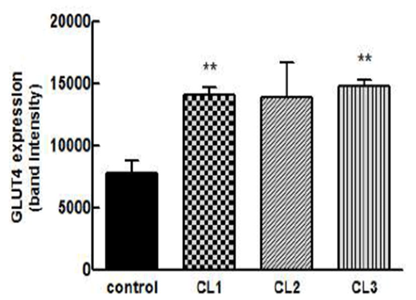
마우스 근육세포에서 CL01 처리에 따른 GLUT4 mRNA 발현양을 확인한 결과(Fig. 1), 근육세포에 CL01을 각각 100, 250, 500 μg/mL로 처리했을 때 GLUT4 유전자가 음성 대조군 대비 각각 약 180%, 178%, 189%(p<0.01) 발현되었 다. 본 연구에서 CL01 처리를 통해 근육세포에서 GLUT4 mRNA 발현이 증가하는 것을 확인하였다.

kjfp-22-6-893-g1
Fig. 1. Effects of CL01 on GLUT4 mRNA expression in C2C12 cell. Control, negative control; CL1, CL01 treated at 100 ㎍/mL; CL2, CL01 treated at 250 ㎍/mL; CL3, CL01 treated at 500 ㎍/mL. Data are expressed as mean±SE (n=3). The results were statistically analyzed by Student’s t-test method. **Significantly different from control (p<0.01).
Download Original Figure
CL01이 체중, 혈당에 미치는 영향

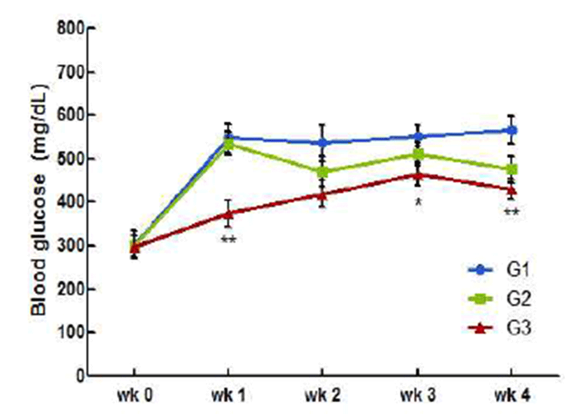
4주간 CL01 투여에 따른 체중 변화를 살펴보기 위해 주 1회 체중을 측정하였다(Table 3). 체중 측정 결과, 모든 실험군에서 유의적인 변화가 관찰되지 않았으며 실험 기간 내내 체중이 증가하는 경향을 보였다. 실험기간 동안의 혈 당 변화를 측정하기 위해 주 1회 체중 측정 후 혈당을 측정 하였다(Fig. 2). 투여 1주차에 CL01 투여군의 혈당이 증가하 여 음성대조군과 혈당 수치가 비슷하지만 2주차부터 혈당 이 감소하여 음성대조군보다 약 12.3% 혈당 수치가 낮게 측정 되었고, 시험 종료 시 양성대조군의 약 110% 수준 (475.40±31.16 mg/dL)으로 감소하였다. 4주간의 혈당 변화 를 관찰한 결과, CL01이 전문 혈당강하제에 비해 속효성은 낮으나 장기 투여 시 전문 혈당강하제의 약 66% 수준의 혈당 강하 효력이 있는 것을 알 수 있었다.

kjfp-22-6-893-g2
Fig. 2. Changes in blood glucose levels in C57BL/KsJ-db/db mice fed diets supplemented with CL01. G1, negative control; G2, CL01 administered at 300 mg/kg; G3, positive control (metformin administered at 300 mg/kg). Data are expressed as mean±SE (n=5). The results were statistically analyzed by Student’s t-test method. **Significantly different from G1 (p<0.01).
Download Original Figure
Table 3. Changes in the bodyweight of C57BL/KsJ-db/db mice fed diets supplemented with CL01
Periods G11) G2 G3
0 week 32.25±0.392) 31.48±0.42 31.45±0.47
1 week 38.09±0.32 36.66±0.67 36.33±0.59
2 weeks 40.48±0.46 38.64±0.82 38.95±0.50
3 weeks 40.46±0.54 39.63±1.05 40.61±0.60
4 weeks 40.72±0.58 41.11±1.15 41.77±0.54
Weight gain 8.47±0.45 9.63±0.43††† 10.32±0.39

1) G1, negative control; G2, CL01 administered at 300 mg/kg; G3, positive control (metformin administered at 300 mg/kg).

2) Data are expressed as mean±SE (n=5). The results were statistically analyzed by Student’s t-test method.

Download Excel Table
CL01이 혈액생화학적 지표에 미치는 영향

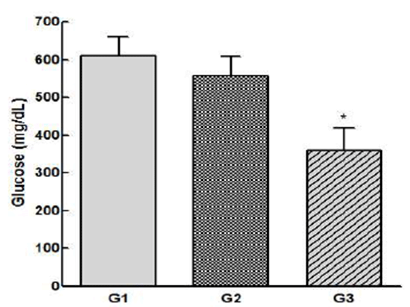
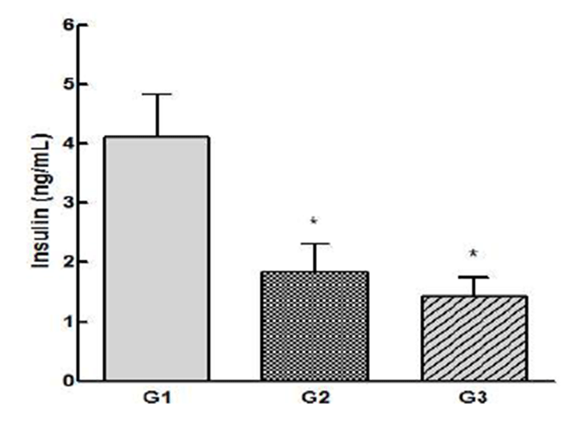
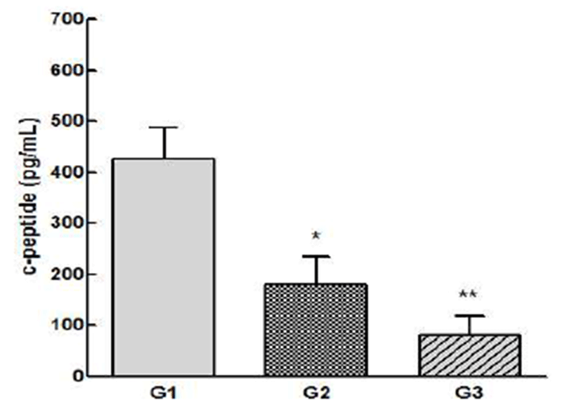
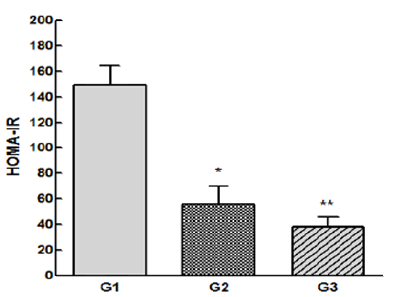
시험 물질 섭취 4주 후 동물을 희생하여 혈장 내 혈당, 인슐린, c-peptide 농도를 측정하고, HOMA-IR 수치를 계산 하였다. 먼저, 혈장 내 혈당 수치를 살펴보면 음성대조군에 비해 양성대조군은 유의적으로 혈당이 감소하였고 (p<0.05), CL01 투여군에서 유의성은 나타나지 않았지만 소폭 감소하였다(Fig. 3). 혈장 내 인슐린 농도를 측정한 결과, 양성대조군의 경우, 음성대조군 대비 약 65% 감소하 여 1.43±0.29 ng/mL(p<0.05)의 농도를 나타냈고, CL01투여 군의 경우, 음성대조군 대비 인슐린 농도가 약 55.8% 감소 하여 1.83±0.47 ng/mL(p<0.05)의 농도를 나타냈다(Fig. 4). 혈장 내 c-peptide 결과를 살펴보면 4주간 시험물질 투약 후 인슐린 농도의 감소와 유사한 형태로 감소한 것을 알 수 있다. 시험 종료 후 양성대조군의 c-peptide 농도는 음성 대조군의 약 20% 수준인 87.56±28.50 pg/mL(p<0.01)의 농 도를 나타냈고, CL01 투여군에서는 음성대조군의 약 40% 수준인 172.69±54.72 pg/mL(p<0.05)의 농도를 나타냈다 (Fig. 5). 분석한 혈액생화학적 지표를 이용하여 인슐린 저 항성을 반영하는 지표인 HOMA-IR 수치를 계산한 결과, 양성대조군에서는 음성대조군 대비 약 74% 인슐린 저항성 이 개선되었고(p<0.01), CL01 투여군에서는 음성대조군 대 비 약 62% 개선되었다(p<0.05)(Fig. 6). 혈장 내 인슐린과 c-peptide 농도는 인슐린 분비의 지표이며, 동시에 인슐린과 c-peptide는 proinsulin의 구성 요소이다. Proinsulin이 분해 되면서 생기는 인슐린은 혈중 안정성이 낮아 분해가 잘 되는 반면 c-peptide는 혈중 안정성이 높아 장시간에 걸친 인슐린 분비능을 확인하는 지표로 활용 가능하다(25). 인슐 린 저항성은 혈당은 동일하되 인슐린 수치가 상이한 서로 다른 개체에서 동일한 혈당을 유지하는데 더 많은 인슐린이 필요하여 그만큼 단위 인슐린의 작용이 떨어졌다는 것을 의미한다(26). 제 2형 당뇨병 환자의 90% 이상에서 인슐린 저항성이 생겨 고인슐린혈증이 유발되고(27), 당내성 장애, 당뇨병, 고혈압, 비만 등의 인슐린 저항성 증후군 유병률이 인슐린의 농도가 증가할수록 증가하며 인슐린 농도가 낮은 경우보다 대사 이상이 생길 확률이 증가한다(28). 본 연구에 서는 CL01의 섭취에 따라 인슐린 저항성이 개선되며 그에 따라 인슐린, c-peptide 및 혈당을 낮추는 것을 알 수 있다.

kjfp-22-6-893-g3
Fig. 3. Effects of CL01 on plasma glucose levels in C57BL/KsJ-db/db mice fed diets supplemented with CL01. G1, negative control; G2, CL01 administered at 300 mg/kg; G3, positive control (metformin administered at 300 mg/kg). Data are expressed as mean±SE (n=5). The results were statistically analyzed by Student’s t-test method. *Significantly different from G1 (p<0.05).
Download Original Figure
kjfp-22-6-893-g4
Fig. 4. Effects of CL01 on plasma insulin in C57BL/KsJ-db/db mice fed diets supplemented with CL01. G1, negative control; G2, CL01 administered at 300 mg/kg; G3, positive control (metformin administered at 300 mg/kg). Data are expressed as mean±SE (n=5). The results were statistically analyzed by Student’s t-test method. *Significantly different from G1 (p<0.05).
Download Original Figure
kjfp-22-6-893-g5
Fig. 5. Effects of CL01 on plasma c-peptide in C57BL/KsJ-db/db mice fed diets supplemented with CL01. G1, negative control; G2, CL01 administered at 300 mg/kg; G3, positive control (metformin administered at 300 mg/kg). Data are expressed as mean±SE (n=5). The results were statistically analyzed by Student’s t-test method. *,**Significantly different from G1 (p<0.05, p<0.01).
Download Original Figure
kjfp-22-6-893-g6
Fig. 6. Effects of CL01 on plasma HOMA-IR in C57BL/KsJ-db/db mice fed diets supplemented with CL01. G1, negative control; G2, CL01 administered at 300 mg/kg; G3, positive control (metformin administered at 300 mg/kg). Data are expressed as mean±SE (n=5). The results were statistically analyzed by Student’s t-test method. *,**Significantly different from G1 (p<0.05, p<0.01).
Download Original Figure
CL01이 포도당 수송체 발현에 미치는 영향

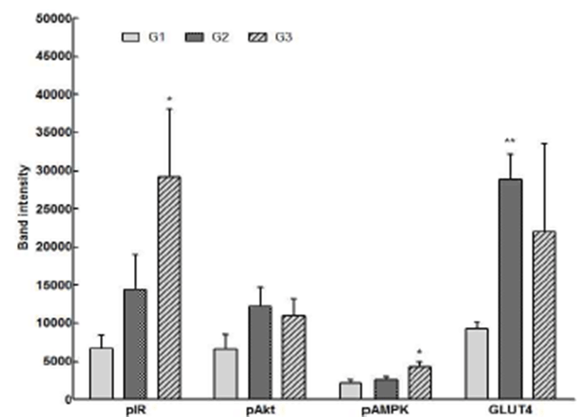
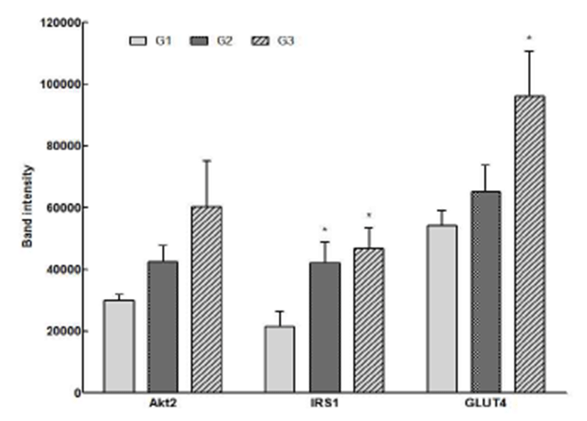
CL01이 근육세포에서 포도당 수송체 발현에 미치는 영 향을 확인하기 위해 시험 물질을 4주간 투여 후 동물을 희생하여 근육세포를 채취하여 포도당 수송체 발현에 관여 하는 유관 단백질 및 mRNA 발현을 측정하였다. Western blot으로 근육세포에서의 단백질을 측정한 결과(Fig. 7), CL01 투여군과 양성대조군에서의 pIR, pAkt, pAMPK 및 GLUT4 단백질 발현이 음성대조군과 비교하여 증가하였 다. 특히 CL01 투여군에서의 pAkt와 GLUT4 단백질 발현양 이 양성대조군 대비 각각 약 10%, 23.6% 증가하였고, pIR와 pAkt 그리고 GLUT4 단백질의 발현양이 음성대조군 대비 각각 약 153%, 145%, 167%(p<0.01) 수준으로 발현되었다. mRNA 발현양을 확인한 결과(Fig. 8), CL01 투여군과 양성 대조군에서의 Akt2, IRS1 및 GLUT4 발현이 음성대조군과 비교하여 증가하였고 CL01 투여군에서의 Akt2와 IRS1이 음성대조군 대비 각각 약 129%, 149%(p<0.05) 발현되었다. 본 연구에서 CL01 섭취를 통해 포도당 수송체 발현과 관련 된 단백질 및 mRNA 발현이 증가하였으며 그에 따라 GLUT4의 발현이 증가하여 혈당 감소에 영향을 미친 것을 확인할 수 있었다. 그리고 CL01 섭취 시 인슐린을 통한 IR의 인산화(pIR)에 도움을 주어 IRS1, Akt2, GLUT4의 유 전자 발현이 연속적으로 이루어져 GLUT4 translocataion을 증가시켜 혈당 감소 효과를 보이며 이는 양성대조군으로 사용한 메트포민과 유사한 기전으로(30), 인슐린에 의한 당 흡수 증가 기전과 다르게 AMPK 활성으로 인한 GLUT4 증가에서 기인한 세포내 당 유입 촉진에 따른 혈당 강하를 확인할 수 있었다. 일반적으로 담자균류에 속하는 버섯의 β-glucan, heteroglyca, galactan 등의 비소화성 수용성 다당 류가 혈당 강하에 우수한 효능을 나타내는 성분으로 알려져 있고(31,32), Kim 등의 연구(33)에서는C. lacerata 균사체 배양 대사산물인 세포외다당체 성분이 인슐린 분비 세포를 덱사메타손의 독성으로부터 보호한다고 보고된 바 있다. 상기의 결과에 비추어 볼 때C. lacerata 균사체 배양 대사산 물인 다당류 등이 항당뇨 활성을 나타내는 것으로 추측할 수 있으며 추후 C. lacerata 균사체 배양물의 기능 성분을 규명하기 위한 기초 증거로 활용할 수 있을 것이다.

kjfp-22-6-893-g7
Fig. 7. Effects of CL01 on protein expression in C57BL/KsJ-db/db mice fed diets supplemented with CL01. G1, negative control; G2, CL01 administered at 300 mg/kg; G3, positive control (metformin administered at 300 mg/kg). Data are expressed as mean±SE (n=5). The results were statistically analyzed by Student’s t-test method. *,**Significantly different from G1 (p<0.05, p<0.01).
Download Original Figure
kjfp-22-6-893-g8
Fig. 8. Effects of CL01 on mRNA expression in C57BL/KsJ-db/db mice fed diets supplemented with CL01. G1, negative control; G2, CL01 administered at 300 mg/kg; G3, positive control (metformin administered at 300 mg/kg). Data are expressed as mean±SE (n=5). The results were statistically analyzed by Student’s t-test method. *Significantly different from G1 (p<0.05).
Download Original Figure

요 약

최근 경제가 급속히 성장하면서 세계적으로 당뇨병이 심각한 대사성 질환으로 대두되고 있으며 이에 따라 혈당 조절에만 초점을 맞춘 경구용 당뇨병 치료제가 아니라 당뇨 병을 근본적으로 치료할 수 있는 혈당 강하 소재를 개발하 기 위한 연구가 늘어나고 있다. 본 연구에서는 선행 연구에 서 항당뇨 효능이 확인된Ceriporia lacerata 균사체 배양물 건조물(CL01)을 제 2형 당뇨병 동물 모델인db/db 마우스에 경구투여하여 CL01이 혈액학적 지표 및 인슐린 신호 전달 기전 상의 유관 단백질과 mRNA 발현에 미치는 영향을 평가하였다. CL01을 4주간 투여했을 때 혈당이 지속적으로 감소하였고, 혈청 인슐린, c-peptide 농도도 각각 음성대조 군의 55.8%, 40% 수준으로 감소하였다(p<0.05). 인슐린 저 항성을 반영하는 지표인 HOMA-IR 수치를 계산한 결과, CL01 투여군에서 인슐린 저항성이 음성대조군 대비 약 62% 개선되었다(p<0.05). 또한 인슐린 신호 전달에 관여하 는 pIR, pAkt, pAMPK, GLUT4 단백질 및 Akt2, IRS1, GLUT4 mRNA 발현이 증가하였다. 이들 결과를 통해 CL01 이 당 대사 및 인슐린 신호 전달에 관여하는 유관 단백질 및 유전자 발현에 영향을 미치며 이에 따라 인슐린 저항성 을 개선하여 혈당을 효과적으로 감소시키는 것을 알 수 있다. 제 2형 당뇨병 환자의 90% 이상이 인슐린 저항성으로 인한 고인슐린혈증, 당내성 장애, 고혈압, 비만 등의 대사 이상이 생길 확률이 증가하기 때문에 CL01의 섭취를 통해 GLUT4 발현을 증가시켜 인슐린 저항성을 낮추는 것이 제 2형 당뇨병의 근본적인 치료에 이용될 수 있을 것으로 사료 되며 이 연구가 당뇨병 치료의 추가적인 기전 연구를 위한 기초 증거로 활용될 수 있을 것이다.

REFERENCES

1.

Daisy P, Saipriya K. Biochemical analysis of Cassia fistula aqueous extract and phytochemically synthesized gold nanoparticles as hypolycemic treatment for diabetes mellitus. Int J Nanomedicine. 2012; 7 p. 1189-1202

2.

Korean Statistical Association Annual report on the cause of death statistics. Korean Statistical Association, Seoul, Korea. 2013.

3.

Beckman JA, Creager MA, Lbby P. Diabetes and atherosclerosis : epidermiology, pathophysiology, and management. JAMA. 2002; 287 p. 2570-2581.

4.

Fonseca V. The role of basal insulin therapy in patients with type 2 diabetes mellitus. Insulin. 2006; 1 p. 51-60

5.

Goldstein BJ. Insulin resistance as the core defect in type 2 diabetes mellitus. J Cardiol. 2002; 90 p. 3G-10G

6.

Hwang KY, Kim YH, Cho YS, Park YS, Lee JY, Kang KD, Kim K, Joo DK, Seong SI. Hypoglycemic effect of fermented soybean culture mixed with mulberry leaves on neonatal streptozotocin-induced diabetic rats. J Korean Soc Food Sci Nutr. 2008; 37 p. 452-458

7.

Chung MJ, Lee YS, Kim BC, Lee SB, Moon TH, Lee SJ, Park KH. The hypoglycemic effects of acarviosine-glucose modulate hepatic and intestinal glucose transporters in vivo. Food Sci Biotechnol. 2006; 15 p. 851-855.

8.

Bell GL, Burant CF, Takeda J, Gould GK. Structure and function of mammalian facilitative sugar transporters. J Biol Chem. 1993; 268 p. 161-164.

9.

Belman JP, Habtemichael EN, Bogan JS. A proteolytic pathway that controls glucose uptake in fat and muscle. Endocr Metab Disord. 2014; 15 p. 55-66

10.

Ishiki M, Klip A. Minireview : recent developments in the regulation of glucose transporter-4 traffic : new signals, locations, and partners. Endocrinology. 2005; 146 p. 5071-5078

11.

Ishikura S, Bilan PJ, Klip A. Rabs 8A and 14 and targets of the insulin-regulated Rab-GAP AS160 regulating GLUT4 traffic in muscle cells. Biochem Biophys Res Commun. 2007; 353 p. 1074-7079

12.

Hayashi T, Wojtaszewski JF, Goodyear LJ. Exercise regulation of glucose transport in skeletal muscle. Am J Physiol. 1997; 273 p. 1039-1051.

13.

Kang HJ. Exercise physiologic mechanisms related to effective exercise prescription in type 2 diabetes mellitus. Korean J Health Promot Dis Prev. 2007; 7 p. 9-16.

14.

Thirone AC, Huang C, Klip A. Tissue-specific roles of IRS proteins in insulin signaling and glucose transport. Trends Endocrinol Metab. 2006; 17 p. 72-78

15.

Thong FS, Dugani CB, Klip A. Turning signals on and off : GLUT4 traffic in the insulin-signaling highway. Physiology (Bethesda). 2005; 20 p. 271-284

16.

White MF. Insulin signaling in health and disease. Science. 2003; 302 p. 1710-1711

17.

Van-pelt RE, Jones PP, Davy KP, Desouza CA, Tanaka H, Davy BM, Seals DR. Regular exercise and the age-related decline in resting metavolic rate in women. J Clin Endocrinol Metab. 1997; 82 p. 3208-3212

18.

Minokoshi Y, Kahn CR, Kahn BB. Tissue-specific ablation of the GLUT4 glucose transporter or the insulin receptor challenges assumptions about insulin action and glucose homeostasis. J Biol Chem. 2003; 278 p. 33609-33612

19.

Lee JW, Gwak KS, Park JY, Park MJ, Choi DH, Kwon M, Choi IG. Biological pretreatment of softwood Pinus densiflora by three white rot fungi. J Microbiol. 2007; 45 p. 485-491.

20.

Lin Y, He X, Han G, Tian Q, Hu W. Removal of crystal violet from aqueous solution using powdered mycelia biomass of Ceriporia lacerata P2. J Environ Sci. 2011; 23 p. 2055-2062

21.

Park JY. Biodegradation of dimethyl phthalate by white rot fungus, Ceriporia lacerata. MS thesis. Seoul National University Seoul Korea. 2007.

22.

Kim JE, Kim HJ, Lee SP. Hyperglycemic effect of submerged culture extract of Ceriporia lacerata in streptozotocin-induced diabetic rats. Food Sci Biotechnol. 2012; 21 p. 1685-1693

23.

Shin EJ, Kim JE, Kim JH, Park YM, Yoon SK, Jang BC, Lee SP, Kim BC. Hypoglycemic effect of submerged culture of Ceriporia lacerata mycelium. Korean J Food Preserv. 2015; 22 p. 145-153

24.

Wannernes F, Caprio M, Gatta L, Fabbri A, Bonini S, Moretti C. Androgen receptor expression during C2C12 skeletal muscle cell line differentiation. Mol Cell Endocrinol. 2008; 292 p. 11-19

25.

Matthews DR, Hosker JP, Rudenski AS, Naylor BA, Treacher DF, Tuner RC. Homeostasis model assessment : insulin resistance and beta-cell function from fasting plasma glucose and insulin concentrations in man. Diabetologia. 1985; 28 p. 412-419.

26.

Jung UJ, Lee MK, Jeong KS, Choi MS. The hypoglycemic effects of hesperidin and naringin are partly mediated by hepatic glucose-regulating enzymes in C57BL/KsJ-db/db mice. J Nutr. 2004; 34 p. 2499-2503

27.

Remsberg KE, Talbott EO, Zborowski JV, Evans RW, McHugh-Pemu K. Evidence for competing effects of body mass, hyperinsulinemia, insulin resistance, and androgens on leptin levels among lean, overweight, and obese women with polycystic ovary syndrome. Fertil Steril. 2002; 78 p. 479-486

28.

Haffner SM, Stern MP, Hazuda HP, Mitchell BD, Patterson JK. Cardiovascular risk factors in confirmed pre-diabetic individuals : does the clock for coronary heart disease start ticking before the onset of clinical diabetes?. JAMA. 1990; 263 p. 2893-2898

29.

Kim JS, Park KS Lee YY. Characteristic of insulin resistance in Korea. Diabetes. 1998; 22 p. 84-91.

30.

Ha JH, Lee SH. Role of AMPK in the regulation of cellular energy metabolism. Endocrinol Metab. 2010; 25 p. 9-17

31.

Yuan Z, He P, Cui J, Takeuchi H. Hypoglycemic effect of water-soluble polysaccharide from Auriculariajudae Quel. on genetically diabetic KK-A mice. Biosci Biotechnol Biochem. 1998; 62 p. 1898-1903.

32.

Kiho T, Sobue S, Ukai S. Structural features and hypoglycemic activities of two polysaccharides from a hot-water extract of Agrocybe cylindracea. Carbohydr Res. 1994; 251 p. 81-87

33.

Kim JH, Park YK, Kim JE, Lee SP, Kim BC, Jang BC. Crude extract of Ceriporia lacerata has a protective effect on dexamethasone-induced cytotoxicity in INS-1 cells via the modulation of PI3K/PKB activity. Int J Mol Med. 2013; 32 p. 179-186

Journal Title Change

We announce that the title of our journal and related information were changed as below from January, 2024.


Before (~2023.12)

After (2024.01~)

Journal Title

Korean Journal of Food Preservation

Food Science and Preservation

Journal Abbreviation

Korean J. Food Preserv.

Food Sci. Preserv.

eISSN

2287-7428

3022-5485

pISSN

1738-7248

3022-5477

Journal Homepage

https://www.ekosfop.or.kr

Same


I don't want to open this window for a day.