1. Introduction
The growing demand for gelatin has driven the search for alternative sources beyond conventional mammalian raw materials, particularly due to halal concerns and sustainability issues. Fish processing by-products, especially skin, represent a promising alternative, with tuna skin being of particular interest due to its high collagen content and wide availability. In addition to addressing religious constraints, the utilization of tuna skin contributes to waste valorization and supports environmentally sustainable production (Karim and Bhat, 2009; Nitsuwat et al., 2021).
However, gelatin derived from fish, including tuna skin, generally exhibits inferior functional properties compared to mammalian gelatin, such as lower gel strength, weaker viscoelasticity, and lower thermal stability. These limitations are mainly attributed to differences in amino acid composition, particularly the lower content of proline and hydroxyproline, which are essential for stabilizing gelatin structure (Derkach et al., 2020; Simon et al., 2003). Therefore, improving the structural and functional properties of tuna skin gelatin remains a key challenge.
Ultrasound-assisted extraction (UAE) has emerged as a promising technique to enhance extraction efficiency and modify protein structure. Through acoustic cavitation, UAE improves mass transfer and disrupts tissue matrices, potentially increasing gelatin yield and altering its molecular conformation (Chemat et al., 2012). Previous studies suggest that UAE can influence protein secondary structure; however, its specific effects on tuna skin gelatin remain insufficiently explored.
Fourier transform infrared spectroscopy (FTIR) is widely used to evaluate protein structural characteristics, particularly through analysis of amide bands that reflect secondary structure changes (Lin et al., 2019). Understanding these structural modifications is essential for linking extraction methods to gelatin functionality.
Therefore, this study aims to investigate the effect of UAE on the chemical structure of tuna skin gelatin using FTIR analysis. This work also evaluates the potential of UAE as a scientific approach to improve the structural quality of gelatin, contributing to the development of halal and sustainable gelatin for industrial applications.
2. Materials and methods
Tuna skin samples were obtained from a frozen tuna export company in Makassar, South Sulawesi, Indonesia. The tuna skins were cleaned to remove scales and residual adhering flesh and then washed with distilled water. The skins were subsequently cut into pieces of approximately 1 cm2 to facilitate the dissolution of gelatin proteins. The size reduction was performed to enhance the solubility of gelatin proteins contained in the tuna skin. The cut samples were placed in polyethylene plastic bags and stored in a freezer at −20° prior to use.
Tuna skin samples were subjected to a sequential pretreatment process to remove non-collagenous proteins and to facilitate collagen swelling before extraction. Initially, the samples were immersed in 0.2 M NaOH (Merck, Darmstadt, Germany) solution at a ratio of 1:6 (w/v) and stirred at 125 rpm for 1 h at room temperature (25°C) to eliminate non-collagen proteins and impurities. After treatment, the alkaline solution was removed, and the samples were thoroughly washed with distilled water until the pH reached neutral. Subsequently, the samples were treated with 0.2 M acetic acid (Merck) at a 1:6 (w/v) ratio under continuous stirring at 125 rpm for 1 h to promote collagen swelling. Following acid treatment, the samples were again washed with distilled water until neutral pH before further extraction processes.
The UAE conditions were selected based on previous studies and the need to balance extraction efficiency and protein structural integrity. Low-frequency ultrasonication (20 kHz) was performed using an Elmasonic S 40 H ultrasonic device (Germany). (Chemat et al., 2012; Kuldiloke, 2002). The ultrasonic power (750 W) was chosen to provide sufficient energy for efficient extraction while minimizing excessive protein degradation (Ahmad et al., 2018). The extraction temperature (55°C) falls within the optimal range for collagen denaturation into gelatin (40-60°C), ensuring effective conversion without significant thermal degradation (Karim and Bhat, 2009). An extraction time of 1 h was applied to maximize yield while avoiding prolonged exposure that may adversely affect gelatin properties. The solvent-to-material ratio (1:6, w/v) was used to ensure adequate solvent penetration and efficient mass transfer during extraction.
This study employed three treatment methods and one control, consisting of: Control (EC), whose conditions (55°C, 18 h, and 300 rpm) were selected based on conventional gelatin extraction protocols. The temperature (55°C) lies within the optimal range for collagen denaturation (45-60°C), ensuring efficient conversion without significant degradation. A longer extraction time (18 h) compensates for the absence of ultrasound, allowing sufficient gelatin diffusion (Alfaro et al., 2015). Stirring (300 rpm) enhances mass and heat transfer. These conditions are widely reported in fish gelatin extraction, including tuna skin, supporting their suitability. Method A: ultrasonic extraction followed by conventional extraction (UAE-EC). Method B: conventional extraction followed by ultrasonic extraction (EC-UAE). Method C: ultrasonic extraction followed by conventional extraction and finalized with ultrasonic extraction (UAE-EC-UAE). The extraction products from the control and Methods A, B, and C were each centrifuged at 6,000 rpm at room temperature for 10 min. The resulting supernatants were then freeze-dried at −50°C under a pressure of 120 mbar for 48 h to obtain gelatin sheets. The gelatin sheets were subsequently ground into gelatin powder. Although Method C involved repeated ultrasonic treatment and therefore a higher cumulative energy input, the treatment sequence was designed to distribute the mechanical effects of ultrasound across two stages rather than applying prolonged ultrasonic exposure continuously. The first ultrasonic step was expected to disrupt the tuna skin matrix and improve solvent penetration, while the conventional extraction step promoted collagen-to-gelatin conversion under thermal conditions. The final ultrasonic treatment was expected to enhance molecular dispersion and extraction efficiency without causing extensive degradation of the main polypeptide structure.
Tuna skin gelatin samples (0.5 g) were weighed and dissolved in 1 mL of distilled water at 60°C. The gelatin solution was placed onto an Attenuated Total Reflectance (ATR) crystal with dimensions of 68 × 8 × 3 mm and analyzed using FTIR spectroscopy. The analysis was performed using a PerkinElmer Spectrum 100 equipped with a single-reflection MIRacle™ ZnSe ATR plate (PerkinElmer, Norwalk, CT, USA).
All samples were scanned over a wavenumber range of 600-4,000 cm−1 at a resolution of 4 cm−1, averaged over 32 scans, and each experimental sample was analyzed in duplicate. ATR-IR images were recorded in the range of 750-4,000 cm−1 with a spectral resolution of 16 cm−1 and a pixel size of 1.56 µm over an area of 300 × 300 μm. The interferometer scanning speed was set at 2.2 cm/s and repeated eight times per pixel. Background spectra were also collected from areas without a protein matrix. The distribution of proteins and fatty acids within the composite was mapped as gradient color spectra, representing characteristic intensity variations for each component.
3. Results and discussion
FTIR analysis was conducted to identify the chemical structure of gelatin produced from tuna skin. The FTIR results revealed characteristic absorption bands corresponding to Amide groups (Amide A, B, I, II, and III), carbonyl (C = O), and hydroxyl (−OH) functional groups, which are the primary indicators in gelatin characterization. The functional group characteristics of tuna skin gelatin extracted using different methods exhibited slight variations in wavenumber values, indicating structural modifications induced by the different extraction treatments (Table 1).
| Catchment area (cm−1) | Wavenumber (cm−1) | Notes | Reference | |||
|---|---|---|---|---|---|---|
| Control | Method A1) | Method B2) | Method C3) | |||
| Amide A 3,300-3,500 | 3,291.14 | 3,290.81 | 3,294.03 | 3,271.71 | Vibration stretching N-H | Muyongga et al. (2004) |
| Amide B 2,915-2,935 | 2,923.17 | 2,922.74 | 2,922.86 | 2,921.13 | Asymmetrical stretching CH2 | Coates (2000) |
| Amide I 1,600-1,690 | 1,639.52 | 1,635.97 | 1,636.48 | 1,635.84 | Vibration stretching C = O | Kong and Yu (2007) |
| Amide II 1,335-1,560 | 1,456.34 | 1,538.76 | 1,542.93 | 1,451.03 | CN stretching, NH bending | Kong and Yu (2007) |
| Amide III 1,229-1,301 | 1,152.71 | 1,237.97 | 1,236.47 | 1,235.93 | CH stretching, NH bending | Kong and Yu (2007) |
The FTIR spectrum of gelatin obtained by conventional extraction (control) showed characteristic absorption bands associated with Amide A, B, I, II, and III structures (Table 1). For instance, the Amide A band was observed at 3,291.14 cm−1, corresponding to N-H stretching vibrations, which indicates good gelatin quality (Muyonga et al., 2004). Comparisons with the literature demonstrated consistency in the Amide A region related to N-H vibrations, as well as in the Amide I band, which corresponds to C = O stretching vibrations at 1,639.52 cm−1 (Kong and Yu, 2007). In the gelatin extracted using the conventional extraction (EC) method, the FTIR spectra showed characteristic absorption bands corresponding to the typical gelatin structure, with no significant shifts observed in the Amide I and III regions, indicating relatively stable secondary structures. In contrast, gelatin obtained using UAE exhibited slight shifts in the wavenumbers of Amide I and III, suggesting alterations in the protein secondary structure. In the UAE-treated samples, the Amide I absorption band shifted to 1,635.97 cm−1, suggesting an increase in β-sheet content, likely due to the mechanical effects of ultrasonic cavitation. These findings are consistent with previous studies reporting that ultrasound treatment can modify the secondary structure of gelatin by reducing α-helix content and increasing β-sheet structures (Ahmad et al., 2018; Wang et al., 2024).
The FTIR spectrum of gelatin obtained by conventional extraction (control) exhibited absorption peaks at 3,291.14 cm−1 (Amide A), 2,923.17 cm−1 (Amide B), 1,639.52 cm−1 (Amide I), 1,456.34 cm−1 (Amide II), and 1,152.71 cm−1 (Amide III) (Fig. 1). These absorption bands indicate a stable gelatin protein structure and good quality, consistent with values reported in previous studies (Kong and Yu, 2007; Muyonga et al., 2004).
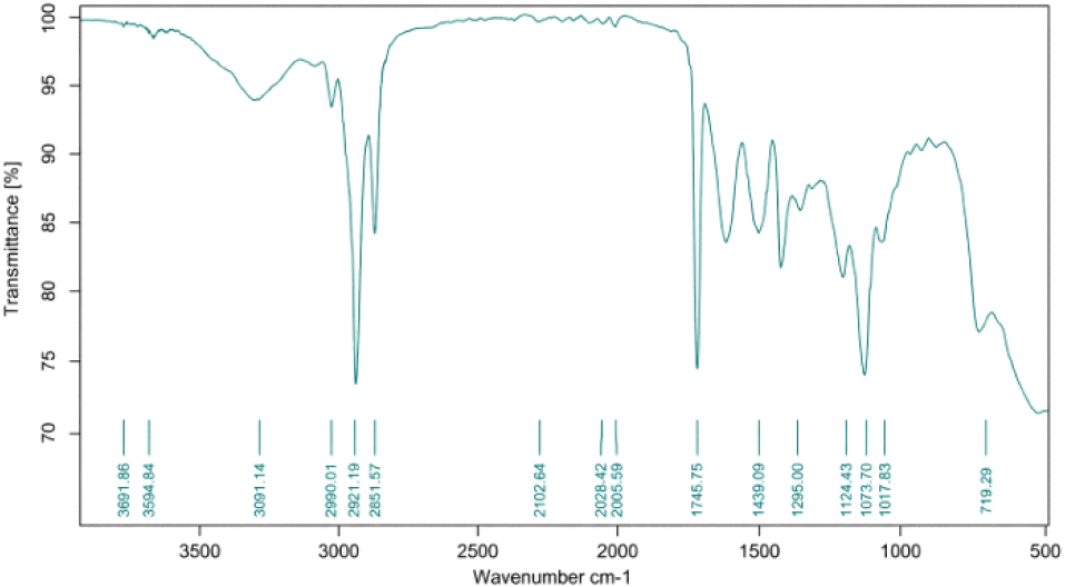
The Amide I absorption band of gelatin derived from tuna skin obtained by conventional extraction (control) was detected at a wavenumber of 1,639.52 cm−1, corresponding to the stretching vibration of the C = O group. Amide I is a characteristic functional group of gelatin. Kong and Yu (2007) reported that Amide I is typically observed in the wavenumber range of 1,600-1,690 cm−1. According to Muyonga et al. (2004), the Amide I band consists of four protein secondary structure components, namely α-helix, β-sheet, β-turn, and random coil structures.
The Amide II absorption band of tuna skin gelatin was detected at 1,456.34 cm−1. Kong and Yu (2007) stated that the Amide II absorption region occurs within the range of 1,560-1,335 cm−1. The presence of the Amide II band indicates C-N stretching and N-H bending vibrations.
As shown in Table 1, the control sample showed an atypical Amide III-related band at 1,152.71 cm−1, which falls outside the typical gelatin Amide III range of 1,229-1,301 cm−1 reported by Kong and Yu (2007).
This value may reflect overlapping C-O/C-N skeletal vibrations, partial peptide-chain disorder, residual non-gelatin components, or incomplete structural reorganization during conventional extraction. Importantly, this anomaly cannot be attributed to thermal treatment alone because Methods A, B, and C were also exposed to 55°C, yet their Amide III bands remained within the expected range (1,235.93-1,237.97 cm−1). Accordingly, the unusually low Amide III-related band in the control sample was considered an anomalous spectral feature, suggesting localized skeletal-vibration irregularity rather than a more ordered or higher-quality gelatin structure.
In contrast, samples obtained using UAE sequences showed Amide III bands within the expected gelatin range. This suggests that ultrasound may have promoted matrix disruption, improved mass transfer, and facilitated more uniform collagen-to-gelatin conversion. Nevertheless, because no non-collagen-treated negative control was included, these FTIR results should be interpreted as evidence of gelatin-associated protein structures and should be supported by complementary analyses in future studies.
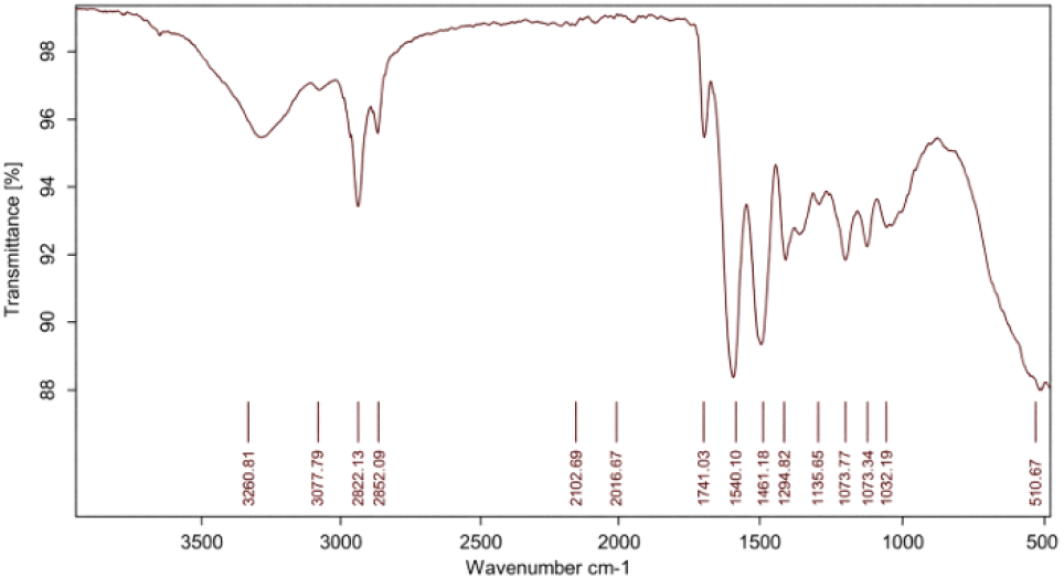
The chemical structure of tuna skin gelatin obtained by ultrasonic-conventional extraction (UAE-EC/Method A) was analyzed using FTIR, which revealed slight shifts in several Amide bands. The Amide A band shifted to 3,290.81 cm−1, the Amide I band to 1,635.97 cm−1, and the Amide III band to 1,237.97 cm−1 (Fig. 2). The shift observed in the Amide III band, for instance at 1,237.97 cm−1, indicates structural changes occurring in the gelatin. These changes are associated with protein structural stability and alterations in hydrogen bonding induced by the extraction method (Stanca et al., 2023). In previous studies, a blue shift in the Amide III band has been linked to the reorganization of the hydrogen-bonding network within protein structures (Derkach et al., 2022). Such shifts suggest that ultrasound treatment induces slight modifications in the secondary structure of gelatin, particularly an increase in β-sheet content and a reduction in α-helix structures, thereby enhancing gelatin flexibility and solubility (Ahmad et al., 2018; Wang et al., 2024).
In Method B (EC-UAE), the Amide A absorption peak was detected at 3,294.03 cm−1, the Amide I band at 1,636.48 cm−1, and the Amide III band at 1,236.47 cm−1 (Fig. 3). The shifts observed in the Amide I and Amide III bands indicate that, although ultrasound was applied, the conventional extraction preceding the ultrasound treatment exerted a greater influence on the gelatin structure.
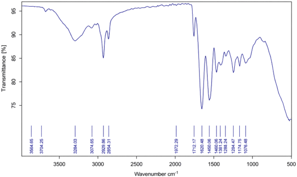
The Amide A band, which is associated with N-H stretching vibrations, confirms the presence of amino and carboxyl groups that play an essential role in the formation of the gelatin gel network. The wavenumbers of the Amide I and Amide III bands detected fall within the standard ranges, representing C = O stretching and N-H bending vibrations of peptide bonds in the protein structure, respectively (Ahmad et al., 2018; Petcharat et al., 2021). The presence and positions of these bands not only confirm the characteristic features of gelatin but also indicate that the structural integrity of the protein was maintained after the extraction process. The slight shifts observed in the Amide I and Amide III bands indicate the influence of extraction conditions, particularly the application of ultrasonic treatment following conventional extraction. Ultrasonication is known to enhance extraction efficiency through cavitation effects that accelerate mass transfer (Usman et al., 2021). However, the results of this study demonstrate that conventional extraction applied at the initial stage plays a dominant role in establishing the basic gelatin structure, while ultrasonic treatment provides only relatively limited additional structural modification (Khawli et al., 2019; Milovanović and Hayes, 2018).
Gelatin obtained using the Method C exhibited characteristic absorption peaks corresponding to Amide A, Amide I, and Amide III. The absorption peaks were observed at 3,271.71 cm−1 for Amide A, 1,635.84 cm−1 for Amide I, and 1,235.93 cm−1 for Amide III (Fig. 4).
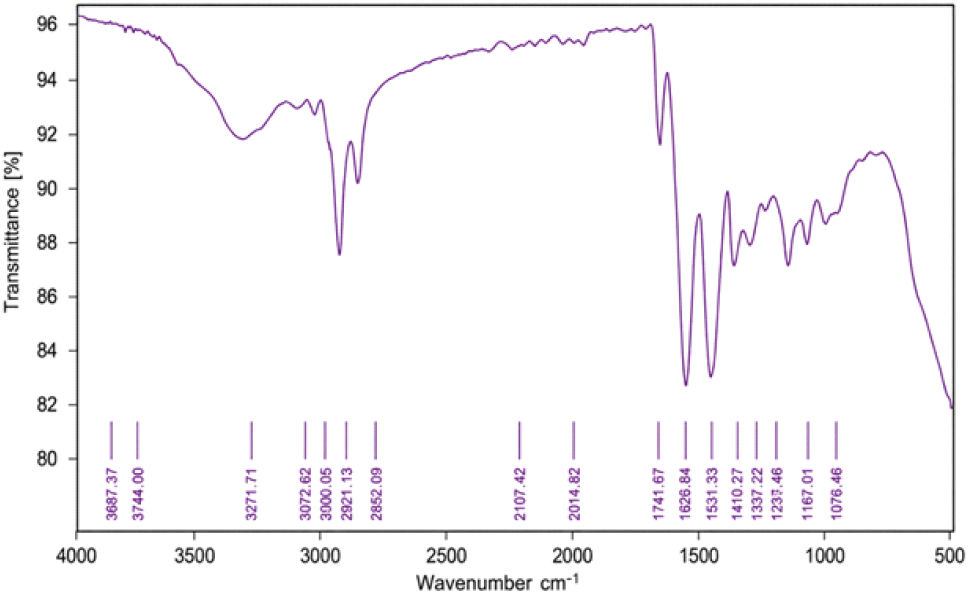
The Amide A absorption peak detected at 3,271.71 cm−1 is associated with N-H stretching vibrations, indicating the presence of hydrogen bonding interactions within the gelatin structure. The Amide A peak commonly observed in gelatin appears within this range, reflecting the structural stability of gelatin during the extraction process (Lima et al., 2025). The results of this study further indicate that the peak at 3,271.71 cm−1 is related to the presence of peptide bonds that are essential for the characteristic properties of gelatin (Sallent et al., 2025). The Amide I band, observed at 1,635.84 cm−1, represents the vibrational mode of the carbonyl (C = O) group within the peptide structure (Sallent et al., 2025). This value is consistent with reports on gelatin derived from various sources, indicating that the extraction method applied did not disrupt the fundamental structure of gelatin (Cuevas-Acuña et al., 2020). This band is considered an important indicator of the presence of triple-helix structures characteristic of gelatin, which remain stable under the conditions investigated (Salinas-Fernandez et al., 2023).
Fig. 5 presents the FTIR spectra of tuna skin gelatin obtained from different extraction treatments: control, Method A (UAE-EC), Method B (EC-UAE), and Method C (UAE-EC-UAE). All samples exhibited the main absorption bands associated with gelatin-like protein structures, particularly in the Amide A, I, II, and III regions. However, the control sample showed an atypical Amide III-related band at 1,152.71 cm−1, while Methods A, B, and C showed Amide III bands within the expected gelatin range.
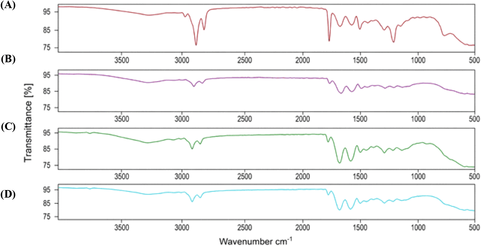
In the Amide A region (3,271-3,294 cm−1), which is associated with N-H stretching and hydrogen bonding, the control and Methods A and B showed similar wavenumbers at approximately 3,290 cm−1, suggesting comparable hydrogen-bonding interactions. Method C exhibited a shift to a lower wavenumber (3,271.71 cm−1), which may indicate stronger hydrogen-bonding interactions after sequential ultrasound-conventional-ultrasound treatment.
The Amide I band (1,635-1,639 cm−1), which reflects protein secondary structure, showed a slight downward shift in all ultrasound-assisted treatments compared with the control. The control sample appeared at 1,639.52 cm−1, while Methods A, B, and C appeared at approximately 1,635-1,636 cm−1. This shift suggests possible secondary-structure rearrangement, including increased beta-sheet contribution and reduced alpha-helix contribution, likely associated with ultrasound-induced molecular reorganization.
For the Amide II region, the control value was 1,456.34 cm−1, while Methods A and B shifted toward higher wavenumbers (1,538.76 and 1,542.93 cm−1, respectively). These shifts may indicate alterations in C-N stretching and N-H bending vibrations due to changes in non-covalent interactions. Method C showed an Amide II band at 1,451.03 cm−1, which was close to the control value and may suggest a relatively stabilized protein conformation after sequential treatment.
In the Amide III region, Methods A, B, and C showed bands at 1,237.97, 1,236.47, and 1,235.93 cm−1, respectively, which fall within the expected gelatin range. In contrast, the control sample showed a markedly lower Amide III-related band at 1,152.71 cm−1. Since Methods A, B, and C were exposed to the same 55°C heating condition and still showed Amide III bands within the expected gelatin range, the anomalous 1,152.71 cm−1 band in the control sample is more likely related to extraction-induced structural variation than to heating alone. It may instead reflect differences in extraction sequence, incomplete structural reorganization, or overlapping spectral contributions in the control sample. Therefore, this control value should be interpreted cautiously as an anomalous spectral feature.
FTIR analysis provides significant insights into the secondary structure of gelatin proteins, particularly through the identification of characteristic bands such as Amide A, Amide I, Amide II, and Amide III. These bands are closely associated with hydrogen bonding, triple-helix conformation, and the integrity of polypeptide chains, which directly contribute to determining the functional properties of gelatin, including viscosity and gel strength.
Gel strength is strongly related to the ability of gelatin to form a three-dimensional (3D) network through the re-association of triple-helix structures, hydrogen bonding, and hydrophobic interactions (Ahmad and Benjakul, 2011; Gómez-Guillén et al., 2011; Karim and Bhat, 2009). The results showed that gelatin extracted from tuna skin using an ultrasonic method exhibited a gel strength ranging from 200 to 220 g (Syahriati et al., 2026). When correlated with the FTIR results, the observed gel strength values did not show any significant shift in the Amide A band (3,271.71-3,290.81 cm−1), indicating that hydrogen bonding remained well preserved. In addition, the clearly detected Amide III band (1,235.93-1,237.97 cm−1) suggests that the helical structure was partially retained. These findings indicate that ultrasonic-assisted extraction of gelatin from tuna skin enhances solvent penetration and accelerates collagen extraction, while preserving the functional protein structure responsible for gel formation.
Gelatin viscosity is influenced by several key factors, including polypeptide chain length, molecular weight distribution, and intermolecular interactions, particularly hydrogen bonding. These characteristics can be analyzed using FTIR, especially through the Amide I (approximately 1,600-1,700 cm−1) and Amide II (approximately 1,500-1,600 cm−1) bands. An FTIR spectrum exhibiting a sharp Amide I band without significant shifts, along with a relatively stable Amide II band, indicates that the secondary protein structure is well preserved and has not undergone excessive degradation. This condition is generally associated with longer and more intact gelatin chains, as well as stronger intermolecular interactions, thereby contributing to increased viscosity.
Based on the results, the viscosity of gelatin extracted from tuna skin using ultrasound ranged from 9 to 10 cP (Syahriati et al., 2026). This finding suggests that ultrasonic treatment (UAE) effectively disrupts the collagen structure without damaging the main polypeptide chains, thereby enhancing the extraction of molecules with relatively higher molecular weight. However, the obtained viscosity values remain below the GMIA standard (15-75 cP), indicating the occurrence of partial chain cleavage (partial hydrolysis). This condition may be reflected in the FTIR spectrum, for instance, through broadening of the Amide I band or a decrease in absorption intensity, which suggests alterations in the protein structure.
4. Conclusions
The FTIR analysis indicated that tuna skin gelatin extracted using ultrasound exhibited gelatin-associated protein spectral features. The Amide A absorption band appeared in the range of 3,271-3,294 cm−1, while the Amide I band at 1,635.84-1,639.52 cm−1 reflected protein secondary-structure characteristics such as alpha-helix and beta-sheet contributions. The Amide III bands of Methods A, B, and C appeared within the expected gelatin range (1,235.93-1,237.97 cm−1), whereas the control sample showed an atypically low Amide III-related band at 1,152.71 cm−1. Overall, the different extraction methods produced broadly similar gelatin-associated spectral characteristics, but the shifts in wavenumber values suggested variations in molecular organization induced by the extraction sequence. Method C (UAE-EC-UAE) showed a relatively stable Amide II and Amide III profile, possibly due to controlled structural reorganization during sequential ultrasound and conventional extraction. However, because this study did not include a non-collagen-treated negative control, the results should be interpreted as FTIR-based evidence of gelatin-associated protein structures rather than absolute confirmation of gelatin purity.