

1. Introduction
Lactic acid bacteria (LAB) play a pivotal role in shelf life extension and microbiological safety in the food industry. LAB produce antimicrobial metabolites through diverse metabolic pathways, including organic acids (primarily lactic acid) and bacteriocins, which lower pH and inhibit spoilage microorganisms and foodborne pathogens (Chen et al., 2025; Zhang et al., 2026). Therefore, natural antimicrobial systems may be effective clean-label preservation strategies that reduce the need for synthetic additives while aligning with consumer demand for minimally processed foods with enhanced storage stability (Marco et al., 2017; Settanni and Corsetti, 2008; Zhang et al., 2026). Additionally, certain LAB strains function as probiotics, conferring health benefits, such as strengthening the intestinal barrier and modulating the host immune system (Azad et al., 2018; Hill et al., 2014). However, for successful application in industrial food systems, candidate strains must exhibit robust technological and safety properties, including tolerance to acidic conditions and bile salts, processing stability, and appropriate antibiotic susceptibility profiles, to ensure functional consistency across diverse environments (Binda et al., 2020; EFSA, 2012; Sanders et al., 2019).
Korea has a well-established tradition of vegetable fermentation, which enhances the seasonal availability and functional properties of agricultural products. Traditional fermented foods, such as kimchi and jang (fermented sauces), are unique ecological niches for autochthonous LAB. These indigenous strains have adapted to harsh environmental conditions, including high osmotic pressures, low temperatures, and complex phytochemical compositions (Jung et al., 2014; Tamang et al., 2016). Compared to commercially standardized starter cultures, these native strains often exhibit greater metabolic flexibility and stress tolerance, making them promising candidates for developing high-value fermented products with extended shelf lives (Di Cagno et al., 2013; Zhang et al., 2026).
Onion (Allium cepa L.), a widely cultivated and economically important crop in Korea, is rich in bioactive compounds such as quercetin glycosides, organosulfur compounds, and fructooligosaccharides (FOS) (Griffiths et al., 2002; Lanzotti, 2006). During LAB-mediated fermentation, quercetin glycosides can be biotransformed into their aglycone forms through microbial β-glucosidase activity, substantially enhancing their bioavailability and functional properties (Filannino et al., 2018; Hur et al., 2014). Additionally, FOS serve as a selective prebiotic substrate that promotes LAB growth and may improve physicochemical stability during storage (Deehan and Walter, 2016). Moreover, organic acids produced during fermentation lower the pH and inhibit spoilage microorganisms, thereby improving shelf stability and potentially reducing the dependence on energy-intensive cold chain systems (Marco et al., 2017; Zhang et al., 2026).
Despite the long-standing consumption of spontaneously fermented onions in traditional Korean diets, systematic investigations on their microbial succession and preservation potential of the dominant LAB remain limited. To address this gap and enhance the value of domestic onion resources, we aimed to isolate and characterize LAB strains from traditional fermented onion products in Korea. The isolates were evaluated for probiotic potential, including gastrointestinal tolerance, adhesion to HT-29 cells, and antimicrobial activity against major foodborne pathogens, as well as key technological and safety properties, such as acid tolerance and antibiotic susceptibility. The novelty of this study lies in the systematic, multi-parameter characterization of the probiotic potential of LAB from traditionally fermented Korean onion (onion jangajji), which has received limited scientific attention as a reservoir of functional LAB despite widespread consumption. To the best of our knowledge, this study is the first comprehensive evaluation of the probiotic properties, safety attributes, and functional characteristics of onion jangajji-derived LAB, providing a scientific basis for identifying candidate strains with multifunctional probiotic potential for further development.
2. Materials and methods
Traditional Korean fermented onions were collected from 10 independent households in Busan and Gyeongsangnam-do, Republic of Korea. Each sample was prepared according to household-specific traditional methods, typically involving an immersion of fresh, sliced onions in a seasoned brine or soy-sauce-based marinade, followed by fermentation at an ambient temperature (15-25°C) for 3-12 weeks. Samples, including both onion tissue and pickling solution, were aseptically collected, transported to the laboratory on ice within 2 h, stored at 4°C upon arrival, and processed within 24 h. For physicochemical analyses, the onion tissue and pickling solution were combined in a 1:1 (w/v) ratio and homogenized using a stomacher (260 rpm, 2 min). For LAB isolation, onion tissue (10 g) and pickling solution (10 mL) were processed separately by diluting each sample in 90 mL of sterile 0.85% NaCl solution.
The pH was measured using a calibrated digital pH meter (Mettler-Toledo, Columbus, OH, USA) following the manufacturer’s instructions. Titratable acidity (TA) was determined by titrating 10 mL of each sample with NaOH (0.1 N) to pH 8.3 and expressed as lactic acid equivalents (%) according to Association of Official Analytical Chemists Official Method 942.15 (AOAC, 2000). Total soluble solids (TSS) were measured using a digital refractometer (PAL-1; ATAGO Co. Ltd., Tokyo, Japan) following the manufacturer’s instructions, and salinity was measured using a digital salinity meter (ES-421; ATAGO Co. Ltd.). The reducing sugar content was determined using the 3,5-dinitrosalicylic acid method with glucose as the standard (Miller, 1959), and absorbance was measured at 540 nm.
Fermented onion samples were serially diluted in sterile 0.85% (w/v) NaCl and spread onto MRS agar supplemented with 1% (w/v) CaCO3. Plates were incubated anaerobically at 37°C for 48-72 h, and colonies exhibiting clear halo formation were selected and purified through two successive subcultures on de Man, Rogosa, and Sharpe (MRS) agar (Difco, BD, Sparks, MD, USA). Presumptive LAB were identified based on gram-positive staining and negative catalase and oxidase activities. Approximately 280 presumptive LAB colonies were recovered from 10 fermented onion samples. Primary screening was conducted by assessing acid tolerance at pH 3.0 for 2 h at 37°C and bile salt tolerance in MRS broth supplemented with 0.3% (w/v) oxgall (Difco) for 4 h at 37°C, as commonly applied in probiotic selection protocols (Charteris et al., 1998). The acid- and bile-tolerance profiles of the recovered isolates were determined, and 74 isolates were subjected to random amplified polymorphic DNA (RAPD)-PCR to assess their genetic relatedness and eliminate potentially redundant isolates. RAPD-PCR was performed using the M13 primer (5′-GAGGGTGGCGGTTCT-3′). RAPD-PCR was performed in a 25-μL reaction mixture containing 50 ng of genomic DNA, 10 pmol primer, 12.5 μL of 2× PCR Master Mix, and nuclease-free water. Amplification was performed with an initial denaturation at 94°C for 5 min, followed by 35 cycles of denaturation at 94°C for 1 min, annealing at 40°C for 1 min, and extension at 72°C for 2 min, with a final extension at 72°C for 10 min. The amplified products were separated using electrophoresis on a 1.5% agarose gel in 1× TAE buffer at 100 V for 60 min, stained with ethidium bromide, and visualized under UV illumination. RAPD fingerprinting patterns were visually compared, and isolates showing ≥ 95% similarity were considered to belong to the same RAPD type. Representative strains were selected from the RAPD groups containing two or more isolates (12 of the 58 groups identified), yielding 12 representative strains (FO-L1 to FO-L12) for 16S rRNA gene sequencing and subsequent probiotic characterization. The remaining 46 RAPD groups, each represented by a single unique isolate, were not selected for further characterization. The RAPD grouping results and representative isolate assignments are summarized in Supplementary Table S1. Genomic DNA of representative isolates was extracted using the DNeasy Blood and Tissue Kit (Qiagen, Hilden, Germany) following the manufacturer’s instructions. The 16S rRNA gene was amplified from ng of genomic DNA using universal primers, 27F and 1492R, in a PCR reaction involving an initial denaturation at 95°C for 5 min, followed by 35 cycles of denaturation at 94°C for 1 min, annealing at 56°C for 1 min, and extension at 72°C for 1 min 30 sec, with a final extension at 72°C for 5 min. The resulting sequences were compared with reference sequences from the National Center for Biotechnology Information GenBank database using the BLAST algorithm. Taxonomic identification was based on ≥ 99% sequence similarity. Phylogenetic analysis was performed using MEGA version 10.0 (Molecular Evolutionary Genetics Analysis, Temple University, Philadelphia, PA, USA), using the neighbor-joining method with 1,000 bootstrap replicates.
Acid tolerance was evaluated following the method described by Charteris et al. (1998) with minor modifications. Bacterial cells were harvested by centrifugation at 7,000 ×g for 10 min at 4°C, washed twice with sterile phosphate-buffered saline (PBS; pH 7.2), resuspended to approximately 1 × 10⁸ CFU/mL in PBS, and adjusted to pH 2.0 and 3.0 using 1 N HCl. Viable cell counts at 0 and 3 h during incubation at 37°C were determined through serial dilution and plating on MRS agar. Bile salt tolerance was assessed by inoculating each strain into MRS broth supplemented with 0.3% and 0.5% (w/v) oxgall and incubating at 37°C. Viable counts were determined at 0 and 4 h. The survival rate (%) was calculated as follows:
where t is the incubation time (3 h for acid tolerance at pH 2.0 and 3.0; 4 h for bile salt tolerance at 0.3% and 0.5% oxgall).
Auto-aggregation was determined as described by Kos et al. (2003). Cell suspensions were prepared in PBS (pH 7.2) to an OD600 of 0.2 (4 mL) and incubated statically at 37°C for 2 h. Auto-aggregation (%) was calculated as follows:
where A0 is the initial absorbacne at 600 nm (0 h) and At is the absorbance at 600 nm of the upper suspension after 2 h of static incubation.
Co-aggregation was evaluated against Listeria monocytogenes KCTC 3569 and Salmonella Typhimurium KCTC 2514 by mixing equal volumes of LAB and pathogen suspensions (OD600 = 0.5) and incubating at 37°C for 2 h. Co-aggregation (%) was calculated as follows:
where ALAB and Apathogen are the absorbances (OD600) of the LAB and pathogen suspensions, respectively, and Amixture is the absorbance (OD600) of the mixed suspension after 2 h of incubation.
Cell surface hydrophobicity was determined using the microbial adhesion to hydrocarbons (MATH) assay with n-hexadecane (Sigma-Aldrich, St. Louis, MO, USA), as described by Rosenberg et al. (1980). Cell suspensions were adjusted to an OD600 of 0.5, and the absorbance at 600 nm was measured before (A0) and after phase separation (A1). Hydrophobicity (%) was calculated as follows:
The human intestinal epithelial cell line, HT-29 (ATCC HTB-38, Manassas, VA, USA), was cultured in Dulbecco’s Modified Eagle Medium (DMEM; Gibco, Thermo Fisher Scientific, Waltham, MA, USA) supplemented with 10% fetal bovine serum (Gibco, Thermo Fisher Scientific) and 1% penicillin-streptomycin at 37°C in a humidified atmosphere containing 5% CO2. Cells were seeded at 1.0 × 10⁵ cells/well in 24-well plates and grown to confluence. HT-29 cells between passages 15 and 25 were used. Cell viability following bacterial exposure was confirmed using the trypan blue exclusion assay (> 95% in all wells). Bacterial suspensions (~1 × 10⁸ CFU/mL in antibiotic-free DMEM) were added at a bacterium-to-cell ratio of approximately 100:1 and incubated for 2 h at 37°C. Lacticaseibacillus rhamnosus GG (LGG, ATCC 53103) was used as a positive control to provide a reference benchmark for the adhesion capacity of a well-characterized probiotic strain. Escherichia coli ATCC 25922, a non-probiotic strain with characteristically low adhesion to intestinal epithelial cells, was used as a negative control to confirm the discriminatory validity of the assay and establish a baseline for non-probiotic adhesion (Tuomola and Salminen, 1998). Both control strains were prepared and inoculated under the identical conditions to those used for the test LAB strains. Non-adherent bacteria were removed by washing three times with PBS (pH 7.2; 1 mL per well, 30 sec incubation before each aspiration). The adherent bacteria were recovered and enumerated by plating. Adhesion (%) was calculated as follows:
where N0 represents the initial number of bacteria added (CFU/well), and N1 represents the number of adhered bacteria (CFU/well).
Antimicrobial activity was evaluated using the agar well diffusion method against L. monocytogenes KCTC 3569 and S. Typhimurium KCTC 2514. Cell-free supernatants (CFSs) were obtained from 24-h cultures in MRS broth using centrifugation at 10,000 ×g for 15 min at 4°C. Crude bacteriocin fractions were prepared by precipitation with 50% saturated ammonium sulfate, followed by dialysis using a dialysis membrane (1 kDa MWCO, Spectra/Por, Spectrum Laboratories, Rancho Dominguez, CA, USA) against 20 mM PBS (pH 6.5) for 24 h at 4°C. Antimicrobial activity was determined by measuring the inhibition zones (mm) after incubation at 37°C for 24 h. The proteinaceous nature of the inhibitory substances was confirmed by treatment with proteinase K (1 mg/mL; Sigma-Aldrich) at 37°C for 2 h. pH-neutralized CFS (pH 6.5) was used as a control to exclude the contribution of organic acids to the observed antimicrobial activity.
Heat stability of the inhibitory substances was assessed by heating pH-neutralized CFS at 80°C for 30 min and 100°C for 20 min in a water bath. After cooling to 25°C, the residual antimicrobial activity was measured against L. monocytogenes KCTC 3569, as described above. To exclude H2O2 as a potential antimicrobial agent, pH-neutralized CFS was treated with catalase (1 mg/mL; Sigma-Aldrich) at 37°C for 1 h, followed by heat inactivation at 80°C for 20 min. A catalase-only control (without CFS) was included to confirm the absence of non-specific enzymatic effects on the indicator strains.
Genomic DNA was extracted using a commercial kit (NucleoSpin Microbial DNA; Macherey-Nagel, Düren, Germany). PCR screening was performed for four representative bacteriocin structural genes: nisin A (nisA), plantaricin A (plnA), sakacin A (sakA), and pediocin PA-1/AcH (pedA), using previously published primer sequences and conditions (Cotter et al., 2013; Khorshidian et al., 2021). PCR products were separated via electrophoresis on 1.5% agarose gels and stained with ethidium bromide.
Hemolytic activity was evaluated by streaking the isolates onto Columbia blood agar (Oxoid, Thermo Fisher Scientific, Basingstoke, UK) supplemented with 5% (v/v) defibrinated sheep blood (Oxoid). Plates were incubated aerobically at 37°C for 48 h. Isolates exhibiting no hemolysis (γ-hemolysis) were considered non-hemolytic, in accordance with safety assessment criteria for LAB (EFSA, 2012).
BA production (histamine, tyramine, putrescine, and cadaverine) was evaluated using decarboxylase screening agar supplemented with the corresponding amino acid precursors (l-histidine, l-tyrosine, l-ornithine, and l-lysine; 1%, w/v; Sigma-Aldrich), pyridoxal-5-phosphate (0.005%, w/v), and bromocresol purple as a pH indicator, according to Bover-Cid and Holzapfel (1999). Plates were incubated at 37°C for 48-72 h, and a color change from yellow to purple indicated a positive reaction.
Virulence-associated genes (gelE, hyl, asa1, cylA, and esp) were detected using PCR with previously validated primers (Franz et al., 2001). Amplification was performed under standard PCR conditions, and the presence of target genes was confirmed using 1.5% (w/v) agarose gel electrophoresis.
Gelatinase activity was phenotypically assessed using MRS agar supplemented with 1.5% (w/v) gelatin. Isolates were spot-inoculated and incubated at 37°C for 24-48 h. Subsequently, plates were flooded with 40% (w/v) ammonium sulfate solution for 15-30 min. Formation of a clear zone surrounding the colonies indicated gelatin hydrolysis and was considered a positive result.
Antibiotic susceptibility was determined by measuring the minimum inhibitory concentrations (MICs) using the broth microdilution method in cation-adjusted Mueller-Hinton broth (Oxoid) supplemented with 5% (v/v) lysed horse blood, according to the Clinical and Laboratory Standards Institute (2020) and European Food Safety Authority (EFSA) 2012 guidelines. Two-fold serial dilutions of ampicillin, penicillin G, erythromycin, tetracycline, chloramphenicol, vancomycin, gentamicin, kanamycin, streptomycin, and clindamycin (Sigma-Aldrich) were prepared in 96-well microplates. After incubation at 37°C for 18-24 h, bacterial growth was determined by measuring absorbance OD600 using a microplate reader (Epoch, BioTek Instruments, Winooski, VT, USA). The MIC was defined as the lowest concentration of antibiotic that completely inhibited visible bacterial growth. The MIC values were interpreted according to the microbiological cut-off values established by EFSA (2012).
Strains that satisfied the predefined probiotic criteria and exhibited acceptable safety profiles were selected for further functional characterization.
Each LAB strain was cultured in MRS broth at 37°C for 18 h and harvested at the stationary phase using centrifugation at 5,000 ×g for 10 min at 4°C. The cell pellets were washed twice with sterile PBS (0.1 M, pH 7.0) and resuspended in PBS to a final OD of 1.0 at 600 nm, corresponding to approximately 108-109 CFU/mL, ensuring comparable cell density across strains prior to each assay. All antioxidant assays were performed using intact cell suspensions.
The DPPH radical-scavenging activity of intact LAB cells was assessed by mixing 1 mL of the cell suspension with 1 mL of 0.2 mM DPPH in 95% ethanol and incubating for 30 min in the dark at 25°C. The mixture was then centrifuged at 5,000 ×g for 10 min, and the absorbance of the supernatant was measured at 517 nm. Scavenging activity was calculated as follows:
where Asample is the abosrobacne of the supernatant of the DPPH-cell mixture at 517 nm and Acontrol is the absorbance of DPPH solution without cells (Brand-Williams et al., 1995).
The ABTS+ radical-scavenging activity was determined by mixing 1 mL of the cell suspension with 1 mL of ABTS+ working solution, prepared by reacting 7 mM ABTS with 2.45 mM potassium persulfate for 16 h in the dark, diluted to an absorbance of 0.70 ± 0.02 at 734 nm. After incubation for 6 min at 25°C, the mixture was centrifuged at 5,000 ×g for 10 min, and the absorbance of the supernatant was measured at 734 nm. Scavenging activity (%) was calculated as follows:
where Asample is the absorbance of the supernatant of the ABTS+-cell mixture at 734 nm and Acontrol is the absorbance of the ABTS+ working solution without cells (Re et al., 1999).
The ferric reducing antioxidant power (FRAP) of intact LAB cells was evaluated by mixing 0.1 mL of the cell suspension with 0.9 mL of freshly prepared FRAP reagent (10 mM TPTZ in 40 mM HCl, 20 mM FeCl3·6H2O, and 300 mM acetate buffer, pH 3.6; 1:1:10, v/v/v) and incubating at 37°C for 10 min. After centrifugation at 5,000 ×g for 10 min, the absorbance of the supernatant was measured at 593 nm, and results were expressed as μmol Fe2+ equivalents per OD600 unit of the standardized cell suspension, using a FeSO4 standard curve (Benzie and Strain, 1996).
Lipid peroxidation inhibitory activity was evaluated using the thiobarbituric acid-reactive substances (TBARS) assay in a linoleic acid emulsion system. An emulsion was prepared by dissolving 0.28 mL of linoleic acid and 0.28 g of Tween 40 in 50 mM PBS (pH 7.0) to a final volume of 50 mL. The cell suspension (1 mL) was mixed with a linoleic acid emulsion (2.5 mL) and incubated at 40°C for 72 h in the dark to induce lipid oxidation. Subsequently, 1 mL of the reaction mixture was combined with 1 mL of 20% trichloroacetic acid and 1 mL of 0.67% thiobarbituric acid (TBA), then heated at 95°C for 30 min. After cooling and centrifugation at 3,000 ×g for 10 min, the absorbance of the malondialdehyde (MDA)-TBA adduct in the supernatant was measured at 532 nm. Inhibitory activity was expressed as a percentage relative to the cell-free control (Ohkawa et al., 1979).
Cholesterol assimilation was evaluated according to Gilliland et al. (1985). MRS broth supplemented with 0.3% (w/v) oxgall and 100 μg/mL cholesterol was inoculated with 1% (v/v) of each strain and incubated anaerobically at 37°C for 24 h. Residual cholesterol was quantified at 510 nm using a commercial enzymatic assay kit (Sigma-Aldrich).
EPS production was initially qualitatively screened on MRS agar supplemented with 5% (w/v) sucrose, followed by quantitative determination in MRS broth containing the same sucrose concentration at 37°C for 24 h. The obtained CFS was mixed with chilled absolute ethanol at a ratio of 1:2 (v/v) and incubated at 4°C overnight to precipitate EPS. The precipitated EPS were collected by centrifugation at 10,000 ×g for 10 min at 4°C, washed twice with absolute ethanol to remove residual impurities, dried, and re-dissolved in distilled water. The total carbohydrate content of the extracted EPS was quantified using the phenol-sulfuric acid method with glucose as the standard, and the absorbance was measured at 490 nm.
Enzymatic activity profiles were determined using the API ZYM system (bioMérieux, Marcy-l’Étoile, France) following the manufacturer’s instructions. Inoculated strips were incubated at 37°C for 4 h, and results were interpreted by adding ZYM A and ZYM B reagents and recording color development on a scale of 0-5.
All experiments were performed in triplicate, and the results are expressed as mean ± SD. Statistical significance was evaluated using one-way analysis of variance, followed by Duncan’s multiple range test (SPSS Statistics v26.0; IBM Corp., Armonk, NY, USA). Pearson’s correlation analysis was performed using the same software. Differences were considered statistically significant at p < 0.05.
3. Results and discussion
The physicochemical characteristics of the fermented onion samples are summarized in Table 1. All 10 fermented onion samples exhibited strongly acidic profiles, consistent with active LAB-mediated fermentation. The pH ranged from 4.24 ± 0.03 (FO-1, 3 weeks) to 3.26 ± 0.10 (FO-10, 12 weeks), with TA increasing correspondingly from 0.55 ± 0.05% to 1.30 ± 0.04% (lactic acid equivalents). The strong inverse correlation between fermentation duration and pH (r = −0.89, p < 0.01) reflects a progressive organic acid accumulation by the resident LAB community. This phenomenon occurs in spontaneously fermented vegetable systems, in which LAB metabolism leads to continuous acid production and concomitant pH decline (Filannino et al., 2018; Hutkins, 2006).
TSS ranged from 12.5 ± 0.8 °Brix (FO-9) to 20.8 ± 0.5 °Brix (FO-10), whereas reducing sugars ranged from 24.5 ± 0.5 to 50.6 ± 2.0 mg/mL, reflecting inter-household variability in raw material composition, preparation practices, and initial substrate concentrations. The persistence of substantial residual reducing sugars, even in long-fermented samples, may be attributed to the gradual hydrolysis of inulin-type fructans by microbial β-fructosidases, which continuously release fermentable mono- and disaccharides (e.g., fructose and glucose) and maintain a residual sugar pool despite ongoing microbial consumption (Filannino et al., 2018). Notably, onions contain substantial levels of fructans that hydrolyze more slowly than simple sugars; therefore, these compounds may serve as sustained carbon reservoirs, supporting prolonged LAB metabolic activity throughout fermentation (Di Cagno et al., 2013; Van Loo et al., 1995).
Salinity ranged from 0.73 ± 0.01% to 1.07 ± 0.03%, markedly lower than the 2-3% NaCl levels typically associated with conventional kimchi fermentation (Hutkins, 2006; Jung et al., 2014). These low-salinity conditions reduce osmotic stress, thereby allowing rapid heterofermentative LAB proliferation, particularly Leuconostoc spp., which are less salt tolerant but metabolically active in the early fermentation stages (Kim et al., 2016). These organisms produce organic acids, CO2, and flavor compounds that contribute to the initial acidification. As fermentation progresses, the combined effects of acid accumulation and gradually increasing osmotic and acidic stress favor acid-tolerant homofermentative LAB, driving microbial succession toward species better adapted to low pH conditions (Lee et al., 2021).
Accordingly, the low-salinity environment observed in the present study may favor the dominance of heterofermentative LAB, contributing to acidification and flavor development. Collectively, the combination of low salinity, high fructan availability, and strong acidification may define a distinct ecological niche in traditionally fermented Korean onions, potentially shaping LAB communities adapted to low salt conditions, high carbohydrate availability, and increased acid stress during fermentation (Filannino et al., 2018; Kim et al., 2016).
Approximately 280 presumptive LAB colonies were recovered from 10 traditional Korean fermented onion (jangajji) samples on CaCO3-MRS agar (Fig. 1), yielding 20-30 colonies per sample. Colonies exhibiting clear zones of CaCO3 dissolution, indicative of acid production, were selected for preliminary characterization. All selected isolates were confirmed to be gram positive, catalase negative, and oxidase negative, consistent with the established phenotypic characteristics of LAB (De Vuyst and Leroy, 2007; Holzapfel et al., 2001).
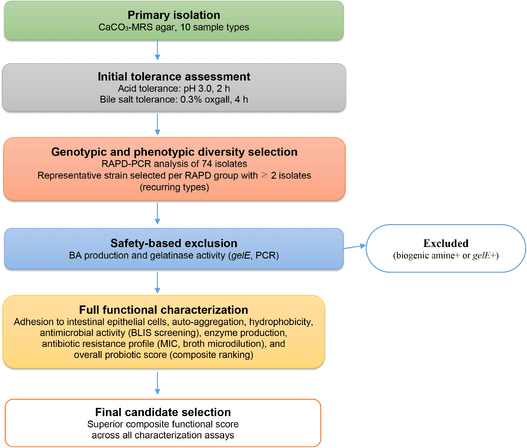
Acid and bile tolerance were assessed as part of the initial characterization of the recovered LAB isolates. Acid tolerance was evaluated after exposure to MRS broth adjusted to pH 3.0 for 2 h at 37°C, whereas bile tolerance was evaluated in MRS broth supplemented with 0.3% (w/v) oxgall for 4 h at 37°C. Seventy-four isolates were subsequently subjected to RAPD-PCR to assess their genetic relatedness and identify potentially redundant isolates.
RAPD fingerprinting patterns were compared, and isolates showing ≥ 95% similarity were considered to belong to the same RAPD type. The 74 isolates were divided into 58 RAPD groups, and representative strains were selected from the groups containing two or more isolates. Twelve representative strains (FO-L1 to FO-L12) were selected for 16S rRNA gene sequencing and comprehensive probiotic characterization, whereas 46 singleton RAPD groups were not selected for further characterization. The RAPD grouping results and representative isolate assignments are summarized in Supplementary Table S1. The overall clustering patterns of the 74 isolates across 58 RAPD groups are shown in Supplementary Fig. S1, and the corresponding per-isolate cluster assignments and selection statuses are listed in Supplementary Table S1. Subsequently, 12 representative strains, designated FO-L1 to FO-L12, were selected for further probiotic evaluation.
Using 16S rRNA gene sequencing, 12 representative strains were identified as 12 species across seven genera (Lactiplantibacillus, Limosilactobacillus, Leuconostoc, Levilactobacillus, Weissella, Pediococcus, and Lacticaseibacillus) with sequence similarities of 99.0-100.0% to reference strains (Table 2), supporting reliable species-level taxonomic assignment (Stackebrandt and Goebel, 1994). The taxonomic composition observed in this study is consistent with previous reports on LAB communities in fermented plant-based foods, in which Lactiplantibacillus, Leuconostoc, Pediococcus, and Levilactobacillus were the dominant genera (Filannino et al., 2018). The detection of key species, including Lactiplantibacillus plantarum, Leuconostoc mesenteroides, Levilactobacillus brevis, and Pediococcus pentosaceus, reflects the typical microbial succession associated with spontaneous vegetable fermentation, in which early heterofermentative species initiate acidification, subsequently favoring more acid-tolerant homofermentative species (Breidt et al., 2012; Lee et al., 2021). The presence of Weissella spp. further highlights the ecological relevance of this genus in vegetable fermentation, whereas that of Limosilactobacillus fermentum and Lacticaseibacillus spp. suggests the metabolic versatility under prolonged fermentation conditions (Di Cagno et al., 2013; Filannino et al., 2018).
Overall, the multi-genus and multi-species LAB community identified in this study, comprising both hetero- and homofermentative or facultative heterofermentative taxa, reflects the complex microbial ecology of spontaneously fermented foods. These findings suggest that traditionally fermented onions are an underexplored reservoir of phylogenetically diverse LAB with potential probiotic functionality. The 12 representative strains selected through sequential functional screening and RAPD-PCR-based dereplication were subsequently subjected to comprehensive in vitro probiotic evaluation, safety assessment, and functional characterization, as described in the following sections. The strain selection procedure is illustrated in Fig. 1.
Acid tolerance is an essential prerequisite for probiotic functionality, as orally ingested bacteria must survive exposure to gastric conditions (pH 1.5-3.5) for periods ranging from 20 min to 3 h (Conway et al., 1987; Marteau et al., 1997). Survival at pH 2.0 (3 h, 37°C) ranged from 22.7 ± 2.6% (Weissella koreensis FO-L11) to 71.0 ± 6.9% (L. rhamnosus FO-L12) (Table 3). At the postprandial pH of 3.0, all 12 strains exhibited ≥ 70% survival, with Weissella confusa FO-L5 (95.3 ± 5.1%) and L. rhamnosus FO-L12 (92.1 ± 4.7%) showing the highest tolerance.
Among all isolates evaluated in this study, L. rhamnosus FO-L12 demonstrated the highest acid tolerance at pH 2.0. The overall survival range of 22.7-71.0% at pH 2.0 is consistent with the substantial strain-dependent variability in acid tolerance widely reported among LAB from fermented plant foods (Charteris et al., 1998; Filannino et al., 2018). These findings suggest that several isolates, particularly L. rhamnosus FO-L12, W. confusa FO-L5, and L. brevis FO-L4, exhibit a relatively favorable gastrointestinal stress tolerance, supporting their potential for probiotic applications.
This variation in acid tolerance may be attributed to multiple cellular adaptation mechanisms, including the maintenance of intracellular pH homeostasis through proton extrusion systems, increased activity of membrane-bound F0F1-ATPases, and alterations in membrane fatty acid composition that reduce proton permeability (Corcoran et al., 2005). Additionally, acid stress responses, such as stress protein synthesis and amino acid decarboxylation pathway activation, can further enhance survival under low pH conditions (Filannino et al., 2018).
Survival under physiological bile salt conditions is an important factor influencing probiotic persistence and colonization in the small intestine (Begley et al., 2006). At 0.3% (w/v) oxgall, survival ranged from 75.5 ± 1.4% (L. fermentum FO-L2) to 98.1 ± 2.5% (L. rhamnosus FO-L12). In 0.5% oxgall, all 12 strains maintained viability above 65%, with L. rhamnosus FO-L12 (90.6 ± 4.8%) and L. brevis FO-L4 (89.9 ± 1.8%) showing the highest tolerance (p < 0.05; Table 3). Bile salt resistance in LAB is largely associated with bile salt hydrolase (BSH) activity, along with adaptive cellular responses such as membrane fatty acid modification and general stress response mechanisms (Begley et al., 2006; Ruiz et al., 2013).
The high survival rate of L. brevis FO-L4 at 0.3% oxgall (97.6 ± 3.4%) may be associated with BSH activity, which contributes to bile tolerance in lactobacilli under bile stress conditions (De Smet et al., 1995). In contrast, moderate bile tolerance of L. fermentum FO-L2 (66.9 ± 2.7% at 0.5% oxgall) is consistent with previous reports indicating that bile tolerance in L. fermentum is highly strain-dependent, whereas other LAB species, such as L. plantarum and L. rhamnosus, often exhibit relatively strong bile resistance, albeit with notable strain-level variability (Hamon et al., 2011; Panicker et al., 2018; Ruiz et al., 2013).
Auto-aggregation, which is often associated with adhesion and colonization potential, ranged from 41.8 ± 3.7% (L. mesenteroides FO-L3) to 65.3 ± 6.7% (W. koreensis FO-L11) (Table 3). Seven strains exhibited auto-aggregation values > 50%, indicating a relatively strong aggregation capacity compared with that of previously reported probiotic strains (Kos et al., 2003). Auto-aggregation is primarily mediated by cell surface-associated components, including adhesins and aggregation-promoting proteins, as well as physicochemical properties, such as cell surface hydrophobicity. Additionally, extracellular polymeric substances and S-layer proteins contribute to intercellular bridging and structural stabilization, whereas electrostatic interactions further modulate the aggregation behavior. These factors collectively influence the ability of LAB to form cell clusters and may contribute to their adhesion and persistence in the gastrointestinal environment (Collado et al., 2008; Kos et al., 2003; Lebeer et al., 2008).
Co-aggregation with L. monocytogenes KCTC 3569 ranged from 26.1 ± 2.2% (L. plantarum FO-L1) to 52.3 ± 1.5% (W. koreensis FO-L11), whereas that with S. Typhimurium KCTC 2514 ranged from 23.5 ± 4.0% (Lactiplantibacillus paraplantarum FO-L8) to 52.1 ± 4.8% (Leu. mesenteroides FO-L3). These values correspond with those of probiotic LAB that co-aggregate with pathogenic bacteria (Collado et al., 2008). Co-aggregation between LAB and pathogenic bacteria is mediated by specific cell-surface interactions, including protein-protein and carbohydrate-protein binding mechanisms, and is an important factor in the competitive exclusion of pathogens from intestinal epithelial adhesion sites (Boris et al., 1998; Handley et al., 1987).
Cell surface hydrophobicity, determined using the MATH assay with n-hexadecane (Rosenberg et al., 1980), ranged from 39.4 ± 2.8% (L. fermentum FO-L2) to 62.4 ± 5.3% (L. rhamnosus FO-L12) (Table 3). Six strains exhibited hydrophobicity values > 50%, indicating relatively high surface hydrophobicity compared to that of previously reported probiotic strains (Vinderola and Reinheimer, 2003). A strong positive correlation was observed between surface hydrophobicity and HT-29 adhesion capacity (Pearson’s r = 0.78, p < 0.01; Table S2), suggesting that hydrophobic interactions may contribute to the initial, non-specific contact between bacterial cells and epithelial surfaces prior to receptor-mediated adhesion (Van Loosdrecht et al., 1990). These interactions facilitate subsequent stable adhesion and colonization, potentially enhancing the persistence of probiotic strains in the gastrointestinal tract (Lebeer et al., 2008; Ouwehand et al., 2002).
Adhesion to intestinal epithelial cells is a key in vitro criterion for probiotic selection because it reflects the potential for transient mucosal colonization, competitive exclusion of pathogens, and host-microbe interactions (Monteagudo-Mera et al., 2019). The positive control LGG exhibited an adhesion rate of 18.2 ± 2.1% to HT-29 cells. In contrast, the negative control E. coli ATCC 25922 showed a markedly lower adhesion rate of 4.1 ± 0.8%, confirming the validity and discriminatory power of the assay (data not shown). Among the 12 test strains, adhesion ranged from 14.7 ± 0.8% (P. pentosaceus FO-L6) to 34.0 ± 2.5% (L. rhamnosus FO-L12), representing values substantially above those observed for the negative control and comparable to or approaching the positive control benchmark. Particularly, L. rhamnosus FO-L12 and Leuconostoc citreum FO-L9 exhibited the highest adhesion capacity among the isolates (p < 0.05), suggesting a strong potential for epithelial interaction and transient mucosal colonization.
The high adhesion observed for FO-L12 was consistent with the well-documented adhesive capacity of L. rhamnosus, which is associated with surface structures, such as SpaCBA pili and EPS, that facilitate specific binding to mucins and intestinal epithelial surface glycoproteins (Kankainen et al., 2009; Lebeer et al., 2008; Lebeer et al., 2012). A positive correlation was observed between cell surface hydrophobicity and HT-29 adhesion across all 12 strains (Pearson’s r = 0.78, p < 0.01), suggesting that hydrophobic interactions contribute to the initial, non-specific contact between bacterial cells and epithelial surfaces (Van Loosdrecht et al., 1990). In contrast, strains with comparatively lower adhesion values, such as L. fermentum FO-L2 (24.0 ± 2.2%), should not be excluded from probiotic consideration solely on this basis, as adhesion to intestinal epithelial cells is a multifactorial trait influenced by diverse surface properties and host-specific factors (Krausová et al., 2019). Overall, probiotic adhesion potential cannot be assessed based on a single parameter and should be evaluated in conjunction with auto-aggregation, surface hydrophobicity, and other functional traits that collectively determine mucosal interaction capacity.
The antimicrobial activities of the CFSs from the 12 LAB isolates against L. monocytogenes KCTC 3569 and S. Typhimurium KCTC 2514 were evaluated under three conditions: untreated CFS, pH-neutralized CFS (pH 6.5), and proteinase K-treated neutralized CFS (Table 4). All isolates exhibited inhibitory activity against both pathogens when untreated CFS was applied, with inhibition zone diameters against L. monocytogenes ranging from 11.2 ± 0.5 mm (FO-L12) to 21.2 ± 0.9 mm (FO-L10) and S. Typhimurium from 10.3 ± 1.3 mm (FO-L9) to 18.2 ± 0.8 mm (FO-L4). Therefore, all tested LAB strains produced diffusible antimicrobial metabolites capable of inhibiting both gram-positive and gram-negative pathogens.
Heat stability: + indicates inhibitory activity retained (≥ 90% of original); (+) indicates partially reduced activity (statistically significant decrease but not complete loss); − indicates activity abolished.
Catalase treatment (1 mg/mL, 37°C, 1 h): − indicates no significant reduction in inhibitory activity, confirming H2O2 is not the primary active agent.
PCR screening for bacteriocin structural genes nisA (nisin A), plnA (plantaricin A), sakA (sakacin A), pedA (pediocin PA-1/AcH). All (−) indicates all four genes negative.
After pH neutralization to 6.5, the antimicrobial activity was completely abolished (ND) in eight strains (FO-L1, FO-L3, FO-L6, FO-L7, FO-L8, FO-L9, FO-L11, and FO-L12) against both indicator organisms, indicating that the primary antimicrobial mechanism in these isolates was attributable to organic acid production. Organic acids, particularly lactic and acetic acids, exert antimicrobial effects through extracellular acidification and intracellular diffusion of the undissociated acid form, leading to cytoplasmic acidification and the disruption of metabolic functions (Theron and Lues, 2011; Wang et al., 2021). In contrast, four strains, L. fermentum FO-L2, L. brevis FO-L4, W. confusa FO-L5, and Pediococcus acidilactici FO-L10, retained measurable inhibitory activity against L. monocytogenes at pH 6.5 (14.8 ± 0.6, 13.1 ± 0.4, 12.2 ± 0.5, and 15.5 ± 0.5 mm, respectively), suggesting the production of pH-stable antimicrobial substances beyond organic acids. Among the four strains, FO-L2, FO-L4, and FO-L5 retained measurable inhibitory activity against S. Typhimurium after pH neutralization (11.7 ± 1.3, 12.5 ± 1.3, and 10.2 ± 0.5 mm, respectively), whereas FO-L10 completely lost activity. Therefore, the pH-stable inhibitory substances produced by FO-L2, FO-L4, and FO-L5 may have broader antimicrobial activity than those produced by FO-L10.
Subsequent treatment of the pH-neutralized CFS with proteinase K completely abolished all residual inhibitory activities of the four active strains (Table 4), confirming the proteinaceous nature of the antimicrobial substances. This sensitivity to proteolytic degradation is a defining characteristic of ribosomally synthesized antimicrobial peptides and consistent with the properties of bacteriocin-like inhibitory substances (BLISs) produced by LAB (Dobson et al., 2012; Simons et al., 2020). Accordingly, FO-L2, FO-L4, FO-L5, and FO-L10 were classified as BLIS producers and subjected to further characterization.
Heat stability testing of the BLIS-containing CFS from the four strains revealed a consistent pattern. Notably, inhibitory activity was fully retained following treatment at 80°C for 30 min, whereas partial but significant reduction in activity was observed after treatment at 100°C for 20 min (Table 4). This moderate thermostability is characteristic of many class II bacteriocins, which typically maintain activity up to 80-100°C, but may undergo partial structural denaturation at higher temperatures (Cotter et al., 2013; Negash and Tsehai, 2020). The observed partial retention of activity at 100°C suggests that the BLISs produced by these strains have relatively robust thermal tolerance, which is advantageous for potential application in heat-processed food systems. In contrast, L. brevis FO-L4 showed complete loss of activity at 100°C, suggesting that the inhibitory substance produced by this strain may be more thermolabile than those produced by the other three strains, potentially due to differences in structural conformation or molecular weight.
Catalase treatment of the pH-neutralized CFS from all four BLIS-producing strains did not significantly reduce antimicrobial activity, confirming that H2O2 is not the primary active agent responsible for the observed inhibition. H2O2 is a known antimicrobial metabolite of LAB, but it is sensitive to pH neutralization and catalase degradation; its elimination in the present study confirms that the inhibitory substances are of a proteinaceous, non-oxidative nature (Simons et al., 2020). The catalase-only control produced no inhibition zones, ruling out non-specific enzyme effects on the indicator strains.
The PCR-based screening of four representative bacteriocin structural genes, nisA, plnA, sakA, and pedA, revealed that none of the four BLIS-producing strains harbored any of the screened genes. The absence of these commonly characterized bacteriocin structural genes indicated that the inhibitory substances produced by FO-L2, FO-L4, FO-L5, and FO-L10 were not encoded by the genes targeted in this study. Similar findings have been reported in studies on LAB isolated from fermented foods, in which functional antimicrobial activity was demonstrated despite the absence of known bacteriocin genes, underscoring the diversity of antimicrobial peptides produced by food-associated LAB (Simons et al., 2020; Singh et al., 2024).
Among the BLIS producers, P. acidilactici FO-L10 showed the highest inhibitory activity against L. monocytogenes at neutral pH (15.5 ± 0.5 mm) and lost all activity against S. Typhimurium after pH neutralization, suggesting a relatively narrow antimicrobial spectrum. Although pedA was not detected using PCR, the inhibitory spectrum and biochemical properties of FO-L10 were broadly consistent with those of pediocin-class BLIS. In contrast, L. fermentum FO-L2, L. brevis FO-L4, and W. confusa FO-L5 retained their inhibitory activities against both indicator organisms after pH neutralization, indicating the presence of pH-stable proteinaceous antimicrobial substances with broader inhibitory spectra. The complete loss of activity after proteinase K treatment suggests that these inhibitory substances are proteinaceous and substantially contribute to the observed antimicrobial activity against both indicator organisms. The detection of BLIS activity in W. confusa FO-L5 is consistent with reported antimicrobial peptide production in Weissella species, although BLIS production in this genus remains strain-dependent and poorly characterized at the molecular level (Singh et al., 2024).
Collectively, the biochemical characterization results, including pH stability, proteinase K sensitivity, heat stability, and catalase insensitivity, consistently supported the classification of inhibitory substances produced by FO-L2, FO-L4, FO-L5, and FO-L10 as BLIS. However, definitive characterization (e.g., peptide sequencing, genome mining, and MIC determination) was not performed in the present study; therefore, these substances were designated as bacteriocin-like inhibitory substances rather than confirmed bacteriocins. Further molecular and structural characterizations are warranted to fully elucidate the identity and mechanism of action of these promising antimicrobial substances.
Prior to functional characterization, all 12 representative strains were subjected to safety screening according to the established probiotic evaluation criteria (EFSA, 2012; FAO/WHO, 2002). BA production was assessed using the decarboxylase broth method, gelatinase activity was evaluated using a gelatin hydrolysis assay with concurrent PCR detection of the gelE gene, and virulence-associated genes and antibiotic susceptibility were examined using multiplex PCR and phenotypic testing, respectively. Four strains were excluded from further consideration: FO-L1, FO-L3, and FO-L8 due to BA production (histamine and/or tyramine) and FO-L6 due to gelE positivity with confirmed gelatinase activity (Table 5). The detailed safety evaluation results for all 12 strains are presented in the following subsections.
γ-hemolysis (non-hemolytic phenotype), indicating the absence of a hemolytic zone on Columbia blood agar supplemented with 5% (v/v) defibrinated sheep blood after incubation at 37°C for 48 h (EFSA BIOHAZ Panel, 2011).
Virulence genes screened included gelE (gelatinase), hyl (hyaluronidase), asa1 (aggregation substance), cylA (cytolysin activator), and esp (enterococcal surface protein).
MIC, minimum inhibitory concentration values are expressed as μg/mL and were determined according to EFSA (2012) guidelines for the assessment of antibiotic susceptibility in lactic acid bacteria.
All isolates exhibited γ-hemolysis on Columbia blood agar supplemented with 5% (v/v) sheep blood after 48 h at 37°C, with no evidence of α- or β-hemolysis. The absence of hemolytic activity is consistent with previous reports describing non-hemolytic phenotypes in LAB isolated from fermented foods (Bindu and Lakshmidevi, 2021). The absence of β-hemolysis is considered an important safety-related characteristic, as hemolytic activity is associated with potential virulence in certain bacterial groups and is therefore evaluated in safety assessment frameworks for food-grade microorganisms (Franz et al., 2011; Giraffa, 2002). Notably, all isolates exhibited a non-hemolytic phenotype, consistent with probiotic safety requirements.
BA formed via amino acid decarboxylation may pose toxicological risks to consumers (EFSA Panel on Biological Hazards (BIOHAZ), 2011; Shalaby, 1996). Nine isolates were negative for all tested amines, whereas L. plantarum FO-L1 produced tyramine, L. mesenteroides FO-L3 produced both histamine and tyramine, and L. paraplantarum FO-L8 produced histamine and cadaverine. Additionally, the histamine-producing capacity of FO-L3 and FO-L8 is a critical safety concern, as histamine accumulation is a major food safety issue in fermented products, and BA-producing strains are typically considered undesirable probiotic candidates (EFSA Panel on Biological Hazards (BIOHAZ), 2011; Ladero et al., 2010). Accordingly, FO-L1, FO-L3, and FO-L8 were excluded from further consideration as probiotic candidates because of their BA-producing potential. BA is formed through the microbial decarboxylation of amino acids mediated by specific decarboxylase enzymes, which are often induced under acidic and stressful conditions as part of the bacterial survival mechanism. This pathway contributes to intracellular pH regulation but can lead to the accumulation of toxic amines in fermented foods (Ladero et al., 2010; Shalaby, 1996).
PCR screening for five virulence-associated genes (gelE, hyl, asa1, cylA, and esp) revealed that 10 of 12 isolates were negative for all targets. These genes are primarily associated with enterococcal pathogenicity and are located on mobile genetic elements, contributing to their potential for horizontal transfer (Eaton and Gasson, 2001; Vankerckhoven et al., 2004).
P. pentosaceus FO-L6 harbored the gelE gene and exhibited phenotypic gelatinase activity. Gelatinase is a well-characterized virulence factor in enterococci involved in extracellular matrix degradation, biofilm formation, and host tissue damage (Hancock and Perego, 2004; Thurlow et al., 2010). Although gelE detection in non-enterococcal LAB is uncommon, its presence warrants caution because of the potential for horizontal gene transfer and dissemination of virulence determinants (Iweriebor et al., 2015).
L. rhamnosus FO-L12 tested positive for an esp-like sequence; however, esp homologs detected outside clinical enterococci may not necessarily confer virulence, as gene presence does not always correlate with expression or functional pathogenicity (Shankar et al., 1999; Upadhyaya et al., 2011). In accordance with EFSA (2012) recommendations, whole-genome sequencing is required to further evaluate the genomic context and functional relevance of virulence-associated genes.
All the isolates were susceptible to ampicillin, penicillin G, and vancomycin, suggesting no acquired resistance to clinically important cell wall-targeting antibiotics (Danielsen and Wind, 2003; Klare et al., 2005). This is a critical safety criterion for probiotic candidates because the absence of transferable resistance to clinically relevant antibiotics reduces the risk of horizontal gene transfer to commensal or pathogenic bacteria within the gastrointestinal tract (Ammor et al., 2007; EFSA, 2012).
Elevated tetracycline MICs were observed in L. brevis FO-L4 (16 μg/mL) and Lacticaseibacillus paracasei FO-L7 (32 μg/mL), exceeding the EFSA-defined epidemiological cut-off values for the respective species (Ammor et al., 2007; EFSA, 2012). These elevated values may be associated with ribosomal protection genes, such as tet(M) or tet(W), which are frequently located on mobile genetic elements, and further genomic confirmation is necessary to assess their potential for horizontal transferability (Ammor et al., 2007; Thumu and Halami, 2012).
All isolates exhibited high MIC values for aminoglycosides (kanamycin and streptomycin), consistent with intrinsic resistance attributable to the absence of an electron transport chain required for active antibiotic uptake (Bryan and Kwan, 1983; Teuber et al., 1999). This intrinsic resistance is chromosomally encoded and considered non-transferable (EFSA, 2012). However, in FO-L4 and FO-L7, the MIC values for kanamycin (128 μg/mL) and streptomycin (256 μg/mL) exceeded the EFSA-defined epidemiological cut-off thresholds for Lactobacillus spp. (kanamycin: 64 μg/mL; streptomycin: 64 μg/mL) by two- to four-fold (EFSA, 2012). Although aminoglycoside resistance in LAB is typically intrinsic and non-transferable, the concurrent elevation of tetracycline MICs in these two strains introduces uncertainty as to whether co-located acquired resistance determinants are also present on the mobile genetic elements. Therefore, whole-genome sequencing is strongly recommended for FO-L4 and FO-L7, not to confirm aminoglycoside resistance but to specifically rule out the co-occurrence of transferable resistance genes before these strains are considered for any probiotic application (EFSA, 2012).
Overall, W. confusa FO-L5, L. citreum FO-L9, and P. acidilactici FO-L10 exhibited the most favorable antibiotic susceptibility profiles, with all MIC values falling below the EFSA-defined epidemiological cut-off thresholds. These results support the suitability of these three strains as probiotic candidates from an antibiotic safety perspective (EFSA BIOHAZ Panel, 2011).
Based on the safety assessment results described in Section 3.4, four strains (FO-L1, FO-L3, FO-L6, and FO-L8) were excluded from the functional characterization. The remaining eight strains were ranked according to their combined probiotic performance to identify the most promising candidates for a comprehensive functional evaluation.
After the safety-based exclusion of BA producers (FO-L1, FO-L3, and FO-L8) and gelE-positive strain FO-L6, the remaining eight candidates were ranked based on their combined probiotic performance. L. fermentum FO-L2 was excluded due to its comparatively lower bile salt tolerance at 0.5% oxgall (66.9 ± 2.7%) and lowest surface hydrophobicity among all candidates (39.4 ± 2.8%), indicating a relatively limited probiotic robustness. W. koreensis FO-L11 was excluded despite exhibiting the highest auto-aggregation (65.3 ± 6.7%), primarily because of its markedly poor acid tolerance at pH 2.0 (22.7 ± 2.6%), the lowest among all 12 isolates, which may severely limit its functional efficacy under physiological gastric conditions. L. rhamnosus FO-L12 was retained for discussion because of its exceptional adhesion and acid tolerance. Its esp-like sequence requires further genomic evaluation. W. confusa FO-L5, L. citreum FO-L9, and P. acidilactici FO-L10 were selected for comprehensive functional characterization based on their combination of probiotic performance, favorable safety profiles, and full susceptibility to clinically relevant antibiotics.
The antioxidant capacities of W. confusa FO-L5, L. citreum FO-L9, and P. acidilactici FO-L10 were evaluated using DPPH and ABTS radical-scavenging, FRAP, and TBARS inhibition assays (Table 6). DPPH and ABTS assays evaluate the ability of strains to scavenge stable free radicals through hydrogen or electron donation, whereas FRAP reflects overall reducing power via Fe3+ to Fe2+ conversion. In contrast, TBARS inhibition has biological relevance because it measures the suppression of lipid peroxidation and MDA formation (Benzie and Strain, 1996; Brand-Williams et al., 1995; Ohkawa et al., 1979; Re et al., 1999).
L. citreum FO-L9 consistently exhibited the highest antioxidant activity across all assays: DPPH (76.8 ± 4.7%), ABTS (68.2 ± 0.9%), FRAP (1.31 ± 0.07 μmol Fe2+ equivalents per OD600 unit), and TBARS inhibition (52.2 ± 0.6%). P. acidilactici FO-L10 showed moderate activity, whereas W. confusa FO-L5 exhibited comparatively lower antioxidant capacity. These differences were statistically significant (p < 0.05), indicating a clear strain-dependent variability.
These findings are consistent with those of previous studies demonstrating the strain-specific antioxidant activities of LAB isolated from fermented foods (Lin and Chang, 2000; Zhou et al., 2019). Particularly, Leuconostoc and Pediococcus species exhibit radical scavenging and reducing activities (Lin and Chang, 2000; Roşca et al., 2024). The superior performance of L. citreum FO-L9 may be associated with its high EPS production (164.2 ± 2.5 mg/L; Table S3), as LAB-derived EPS have been reported to contribute to antioxidant capacity (Yang et al., 2021; Zhou et al., 2019).
The antioxidant activity of LAB is attributed to multiple mechanisms, including enzymatic detoxification of reactive oxygen species, metal ion chelation, and bioactive metabolite production, such as EPS and fermentation-derived antioxidant compounds (Lin and Yen, 1999; Zhou et al., 2019). The relatively high TBARS inhibition observed with L. citreum FO-L9 further suggested its potential to suppress lipid peroxidation and may support its applicability in oxidative stress-associated conditions.
All the three strains assimilated cholesterol in vitro. L. citreum FO-L9 exhibited the highest cholesterol-lowering activity (40.6 ± 1.3%), followed by P. acidilactici FO-L10 (33.7 ± 0.9%) and W. confusa FO-L5 (26.6 ± 2.0%) (Table 6). These differences were statistically significant (p < 0.05), indicating a clear strain-dependent variability in cholesterol assimilation capacity.
The cholesterol-lowering capacity of probiotic LAB is typically attributed to multiple mechanisms, including BSH activity, which deconjugates bile acids and promotes the co-precipitation of cholesterol in the intestinal lumen, physical adsorption of cholesterol onto the bacterial cell surface, and incorporation of cholesterol into the bacterial cell membrane during growth (Gilliland et al., 1985; Liong and Shah, 2005). Additionally, bile-induced alterations in the membrane lipid composition may further enhance cholesterol uptake and assimilation efficiency.
The relatively high cholesterol assimilation observed for P. acidilactici FO-L10 may be associated with strain-specific physiological and metabolic characteristics, consistent with previous reports of cholesterol-reducing activity in LAB (Lye et al., 2010). Conversely, the lower activity observed in W. confusa FO-L5 compared with that in FO-L9 reflects the well-documented strain-dependent variability in cholesterol assimilation. This variability may also be influenced by culture conditions, such as bile concentration and growth medium composition, which significantly affect cholesterol uptake and metabolism in LAB (Liong and Shah, 2005; Lye et al., 2010).
All three strains produced EPS, as evidenced by mucoid colony formation on sucrose-supplemented MRS agar (Table 6). L. citreum FO-L9 exhibited the highest EPS yield (164.2 ± 2.5 mg/L), followed by P. acidilactici FO-L10 (138.5 ± 3.3 mg/L) and W. confusa FO-L5 (89.4 ± 1.4 mg/L), with statistically significant differences among all strains (p < 0.05).
L. citreum is widely recognized as a prolific EPS producer in fermented food systems, particularly through dextran synthesis, which contributes to texture formation in sourdough and vegetable fermentations (Roşca et al., 2024). Among Weissella species, EPS production was associated with homopolysaccharides exhibiting antioxidant, prebiotic, and techno-functional properties, largely attributed to hydroxyl- and sugar-rich structures capable of electron donation and metal ion interactions (Adesulu-Dahunsi et al., 2018; Kavitake et al., 2020). Similarly, EPS produced by P. acidilactici are associated with functional properties, including antioxidant activity and host-microbe interaction modulation (Song et al., 2021).
Mechanistically, LAB-derived EPS contribute to multiple biological functions through free radical scavenging via hydroxyl group-mediated electron donation (Yang et al., 2021; Zhou et al., 2019), cholesterol reduction through bile acid interactions and micelle disruption (Liong and Shah, 2005), and prebiotic effects by selectively stimulating beneficial gut microbiota and short-chain fatty acid production. Additionally, EPS may enhance the intestinal barrier integrity by promoting mucus layer stabilization and modulating epithelial immune responses (Prete et al., 2021).
Overall, EPS production levels observed in the present study suggest that L. citreum FO-L9 may have enhanced techno-functional and probiotic potential, warranting further structural and functional characterization.
Furthermore, a positive association between EPS production and antioxidant activity was observed in the three selected LAB strains. L. citreum FO-L9, which exhibited the highest EPS yield, also demonstrated the strongest antioxidant activity in DPPH, ABTS, FRAP, and TBARS assays. In contrast, W. confusa FO-L5 produced the lowest amount of EPS and showed a comparatively weaker antioxidant activity, whereas P. acidilactici FO-L10 displayed intermediate levels of both properties.
Although these observations suggested a possible relationship between EPS production and antioxidant activity, the findings should be interpreted with caution, as only three strains were included in the analysis. The Pearson’s correlation coefficients between EPS yield and antioxidant activity indices were high (r = 0.959-0.999); however, these values are presented only for descriptive purposes and their statistical significance cannot be reliably established with this limited sample size. Therefore, the present data do not support a definitive conclusion regarding the relationship between EPS production and antioxidant activity. However, the observed trend is consistent with previous studies reporting that LAB-derived EPS may contribute to antioxidant functionality through free radical scavenging, electron donation, and metal ion chelation mechanisms (Yang et al., 2021; Zhou et al., 2019). Future studies with several strains and detailed structural characterization of EPS are essential to verify this potential association.
Enzymatic activity profiles were evaluated using the API ZYM system (Table 6). Notably, none of the strains exhibited β-glucuronidase activity, indicating a favorable safety profile, as this enzyme is associated with the reactivation of carcinogenic compounds in the colon (Hillege et al., 2024; Kim and Jin, 2001). L. citreum FO-L9 exhibited the highest overall enzymatic activity, showing strong activity (score 4; 20-40 nmol hydrolyzed substrate) for alkaline phosphatase, leucine arylamidase, acid phosphatase, naphthol-AS-BI-phosphohydrolase, β-galactosidase, and α-glucosidase. P. acidilactici FO-L10 showed intermediate enzymatic activity, whereas W. confusa FO-L5 displayed comparatively lower enzymatic activity across most substrates.
These enzymes are associated with key probiotic-relevant functions, including intestinal detoxification (alkaline phosphatase), protein and peptide degradation (leucine arylamidase), phosphate metabolism (acid phosphatase and phosphohydrolase), and carbohydrate utilization (α-glucosidase), collectively contributing to improved nutrient availability and metabolic activity in the gut environment (Salminen et al., 1998). The presence of β-galactosidase activity in all strains may support lactose hydrolysis and improved digestibility in lactose-intolerant individuals (Wang et al., 2021), whereas β-glucosidase activity in FO-L9 suggests the capacity for biotransforming glycosylated phytochemicals into more bioavailable aglycone forms (Filannino et al., 2018). Collectively, the enzymatic profiles of the isolates reflect a combination of safety-relevant and functionally relevant metabolic traits, thereby supporting their potential as probiotic candidates (Bindu and Lakshmidevi, 2021).
4. Conclusions
L. citreum FO-L9 consistently exhibited the most pronounced functional properties across multiple parameters, including EPS production, antioxidant activity, cholesterol assimilation, and enzymatic activity. Furthermore, FO-L9 demonstrated favorable safety characteristics, including the absence of β-glucuronidase activity and susceptibility to clinically relevant antibiotics, supporting its suitability as a probiotic candidate. The combined presence of a high EPS yield, strong bio-functional activities, and metabolic versatility suggests multifunctional probiotic potential of FO-L9 beyond gastrointestinal survival. Notably, L. rhamnosus FO-L12 also demonstrated superior acid tolerance, high HT-29 cell adhesion, and broad-spectrum antimicrobial activity, indicating its outstanding functional properties. However, given the detection of an esp-like sequence, FO-L12 was not selected as a final probiotic candidate, and its suitability for probiotic applications requires further genomic safety evaluation, including whole-genome sequencing, as recommended by EFSA (2012). Collectively, W. confusa FO-L5, L. citreum FO-L9, and P. acidilactici FO-L10 emerged as promising probiotic candidates from traditionally fermented Korean onions, meeting all safety screening criteria and demonstrating multifunctional probiotic properties. Future studies involving in vivo validation, whole-genome characterization, and fermentation optimization are essential to establish the safety, functionality, and industrial applicability of these strains.


